- Clinical Technology
- Adult Immunization
- Hepatology
- Pediatric Immunization
- Screening
- Psychiatry
- Allergy
- Women's Health
- Cardiology
- Pediatrics
- Dermatology
- Endocrinology
- Pain Management
- Gastroenterology
- Infectious Disease
- Obesity Medicine
- Rheumatology
- Nephrology
- Neurology
- Pulmonology
What is causing this woman’s dry cough?
A 39-year-old woman presented with dry cough, which she had had for 3 months. She had mild intermittent asthma and a 5 pack-year smoking history. Her symptoms started after an upper respiratory tract infection and persisted despite multiple courses of antibiotics, decongestants, and corticosteroids.
A 39-year-old woman presented with dry cough, which she had had for 3 months. She had mild intermittent asthma and a 5 pack-year smoking history. Her symptoms started after an upper respiratory tract infection and persisted despite multiple courses of antibiotics, decongestants, and corticosteroids.
The patient experienced episodes of pleuritic chest pain, shortness of breath with exertion, and generalized malaise. This was accompanied by low-grade fevers and night sweats. She reported losing 10 lb during this time. She denied recent history of any sick contacts or travel and had no known exposure to allergens or toxic fumes, but she had had a pet dog and a turtle for many years. Her mother had died of lung cancer at 39 years of age.
Her physical examination findings were remarkable for crackles in the left upper zone. Complete blood cell count revealed leukocytosis (leukocyte count, 20,500/µL), anemia (hemoglobin level, 8.5 g/dL), and thrombocytosis (platelet count, 608,000/μL), and her erythrocyte sedimentation rate was elevated. HIV and purified protein derivative test results were negative, and the sputum specimen was negative for acid-fast bacilli.
The patient's chest radiograph (Figure 1) and CT scan of the chest (Figure 2) are shown.
What is the likely diagnosis?Answer on next page.
ANSWER:
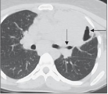
The patient's chest radiograph showed a large left suprahilar mass with cavitation (Figure 1). This was confirmed by a CT scan that showed a large area of consolidation, with an air bronchogram in the left upper lobe and an air-fluid level in the lateral part of the consolidation (Figure 2).
Figure 1 –A large left suprahilar mass is shown in this chest radiograph. The arrow indicates the cavitation.

Figure 2 – This CT scan shows a large area of consolidation, with an air bronchogram in the left upper lobe (arrow) and an air-fluid level in the lateral part of the consolidation (arrow).
An underlying malignancy was suspected on the basis of the radiological findings, the nonresolution despite antibiotic therapy, the patient's smoking history, and her systemic symptoms. Bronchoscopy, transbronchial biopsy, and bronchoalveolar lavage (BAL) were performed. The BAL fluid and the tissue from the biopsy were negative for Mycobacterium tuberculosis, and stains and cultures were negative for fungi.
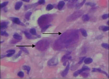
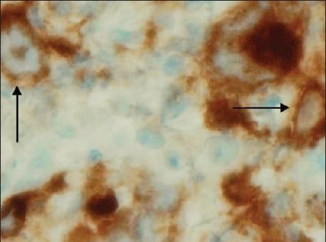
The transbronchial biopsy specimen revealed 2 large atypical cells, which were mildly crushed but had bilobed nucleoli and prominent eosinophilic inclusion bodies-similar to the nucleoli of Reed-Sternberg cells-in a background of small lymphocytes and histiocytes (Figure 3). The presence of Reed-Sternberg cells was confirmed by immunohistological staining that was positive for CD15 and CD30 (Figure 4).

Figure 3 – Two large atypical cells can be seen in this transbronchial biopsy specimen (arrows). The cells are mildly crushed but have bilobed nucleoli and prominent eosinophilic inclusion bodies in a background of small lymphocytes and histiocytes (hematoxylin-eosin stain).

Figure 4 – Immunohistological staining was positive for CD15 and CD30, confirming the presence of Reed-Sternberg cells (arrow).
Additional immunological testing was done to rule out other causes, such as large-cell lymphoma (CD45, CD20) and malignancy (cytokeratin, activin receptor-like kinase-1). The results were negative, thus supporting a definitive diagnosis of Hodgkin lymphoma. The patient had cavitating pulmonary Hodgkin disease.
Discussion
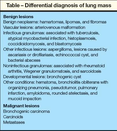
There are many possible causes of a mass or nonresolving infiltrate on a chest radiograph (Table). These disorders must be considered in the differential diagnosis of patients presenting with a pulmonary mass. There also are numerous reasons for the development of cavitating lesions in the lung, including infections (bacterial, tuberculous, nontuberculous mycobacterial, fungal); cystic fibrosis; thromboembolic, vasculitic, and rheumatological diseases; sarcoidosis; silicosis; immunodeficiency; trauma; developmental disorders; and primary and metastatic tumors.1

Pulmonary involvement in Hodgkin disease was first described in 1918 by Walton.2 According to Radin,3 about 15% to 40% of patients with Hodgkin disease will have pulmonary involvement at some time during the course of their disease. It arises in the unencapsulated lymphoid tissue in the lung parenchyma, as well as centrally in the bronchial walls. Connective tissue and subpleural and intraparenchymal lymph nodes also may be involved. Cavitation occurs in fewer than 1% of cases.4 The nodular sclerosing type is the most common histological type.5
In rare cases, primary pulmonary Hodgkin disease can cause a lung mass.6,7 The diagnosis requires the isolated involvement of the pulmonary parenchyma with minimal or no enlargement of the mediastinal and hilar lymph nodes. This is in contrast to our patient, who had involvement of mediastinal lymph nodes on presentation. Only about 100 cases of primary pulmonary Hodgkin disease have been reported in the literature.
Most patients with lung lymphomas present with pulmonary symptoms-most commonly, dry cough. In cases of endobronchial involvement, the cough may become mucopurulent and associated with hemoptysis, although this is unusual. Shortness of breath on exertion may be present. Patients may have chest discomfort that is sharp and pleuritic. One case report described alcohol-induced chest pain.8 Isolated case reports also describe increased itching as a symptom.9 Patients may present with systemic symptoms, such as fever, night sweats, and weight loss. The physical examination may reveal consolidation, wheezing, clubbing, rash, and edema.
In most cases, symptoms occur for months before a diagnosis is made. However, some patients may be asymptomatic with incidental radiological findings.
Lymphomas may appear on a plain chest radiograph as consolidation or cavitation; often, they appear as pneumonic infiltrates that do not resolve with antibiotics. Radiographs may show a solitary mass or multinodular disease. The upper lobes are the most common location.3 Early signs seen on CT scans include diffuse peribronchial thickening. Isolated case reports suggest that CT scans may show an air bronchogram,5,8 as in our patient.
Our patient was treated with doxorubicin, bleomycin, vinblastine, and dacarbazine, and her symptoms improved dramatically.
In conclusion, it is important to recognize pulmonary Hodgkin disease and to differentiate it from other diseases, especially considering its relatively good prognosis.
The authors are affiliated with the department of pulmonary medicine at North Shore University Hospital, Manhasset, New York.
References:
REFERENCES
1.
Ray S, Karbowitz SR. Cavitating pulmonary Hodgkin's disease.
Chest.
2005;128(suppl 4):411S-412S.
2.
Walton HJ. Roentgenological examination of the mediastinum.
AJR.
1918;5:181-185.
3.
Radin AI. Primary pulmonary Hodgkin's disease.
Cancer.
1990;65:550-563.
4.
Horak E, Olinsky A, Chow CW, et al. Multiple cavitating pulmonary nodules and clubbing in 12-year-old girl.
Pediatr Pulmonol.
2002;34:147-149.
5.
Yousem SA, Weiss LM, Colby TV. Primary pulmonary Hodgkin's disease. A clinicopathologic study of 15 cases.
Cancer.
1986;57:1217-1224.
6.
Kern WH, Crepeau AG, Jones JC. Primary Hodgkin's disease of the lung. Report of 4 cases and review of the literature.
Cancer.
1961;14:1151-1165.
7.
Tillawi IS. Primary pulmonary Hodgkin's lymphoma. A report of 2 cases and review of the literature.
Saudi Med J.
2007;28:943-948.
8.
Pik A, Cohen N, Weissgarten J, et al. Primary pulmonary Hodgkin's disease with air bronchogram.
Respiration.
1986;50:226-229.
9.
Dhingra HK, Flance IJ. Cavitary primary pulmonary Hodgkin's disease presenting as pruritus.
Chest.
1970;58:71-73.
