- Clinical Technology
- Adult Immunization
- Hepatology
- Pediatric Immunization
- Screening
- Psychiatry
- Allergy
- Women's Health
- Cardiology
- Pediatrics
- Dermatology
- Endocrinology
- Pain Management
- Gastroenterology
- Infectious Disease
- Obesity Medicine
- Rheumatology
- Nephrology
- Neurology
- Pulmonology
A patient with Cushing syndrome and reduced lung volumes
We present a rare case ofCushing syndrome resultingfrom thymic carcinoid of thelung. Although Cushing syndromeis not usually associatedwith respiratory muscleweakness or restriction, ourpatient had reduced lung volumesand expiratory muscleweakness. His reduced lungvolumes could not be completelyexplained by respiratorymuscle weakness, parenchymallung disease, or obesity.Six months after removal ofthe carcinoid tumor, the patient'sgrowth hormone leveland the lung volumes improvedsignificantly, and hebecame asymptomatic.
We present a rare case of Cushing syndrome resulting from thymic carcinoid of the lung. Although Cushing syndrome is not usually associated with respiratory muscle weakness or restriction, our patient had reduced lung volumes and expiratory muscle weakness. His reduced lung volumes could not be completely explained by respiratory muscle weakness, parenchymal lung disease, or obesity. Six months after removal of the carcinoid tumor, the patient's growth hormone level and the lung volumes improved significantly, and he became asymptomatic.
The case
A 27-year-old nonsmoker was admitted to the hospital complaining of low back pain, weight gain, and proximal muscles weakness for several months. He had cushingoid appearance and proximal muscle weakness in all extremities, especially the quadriceps. A high random urinary free cortisol level led to the diagnosis of Cushing syndrome.
Further workup showed a high serum cortisol level (40 µg/dL [normal, 5 to 25 µg/dL]) and nonsuppressible elevation of adrenocorticotropic hormone (ACTH) in the serum (143 pg/mL [normal, 7 to 69 pg/mL]). The insulin like growth factor I (IGF-I) level, however, was significantly reduced (62 ng/mL [normal, 117 to 329 ng/mL]). No tumor was identified on sella imaging, and findings from bilateral venous sampling of the inferior petrosal sinuses were nondiagnostic.
Findings on a CT scan of the chest and abdomen were normal except for compression deformities of the vertebral bodies of the thoracic and lumbar spine. Therefore, ectopic ACTH production was suspected. An octreotide scan revealed2 areas of abnormality in the chest, consistent with somatastatin receptor–positive neoplasm (Figure 1). No endobronchial lesion was noted on bronchoscopy.
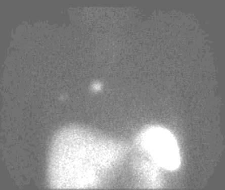
Figure 1 –This octreotide scan of the chest reveals 2 areas of enhancement on the right side, consistent with somatastatin receptor–positive neoplasm.
Preoperative pulmonary function tests revealed the following: forced vital capacity (FVC), 2.5 L (48% of predicted); total lung capacity (TLC), 4.3 L (65% of predicted); vital capacity, 2.9 L (56% of predicted); carbon monoxide–diffusing capacity (DLCO), 19 mL/min/mm Hg (57%) of predicted; and DLCO per unit of alveolar volume, 4.9 mL/min/mm Hg/L (101%) of predicted. These test results were consistent with restrictive lung disease (Table).
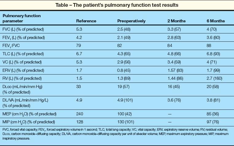
On assessment of the patient's respiratory muscle forces, maximal inspiratory pressure (MIP) was 130 cm H2O (101% of predicted) and maximal expiratory pressure (MEP) was 100 cm H2O (42% of predicted). This suggested expiratory muscle weakness.
The patient then underwent right thoracotomy and wedge resection of the ectopic lesions using the nuclear medicine detection device. The histopathology showed an ACTH producing thymic carcinoid (Figure 2). Pulmonary function tests repeated 2 months later demonstrated that FVC and TLC had increased by 800 mL and 500 mL, respectively. A repeated CT scan of the thoracic and lumbar spine, however, demonstrated osteopenia, with partial collapse of T4, T6, T7, T8, and T9, and complete collapse of T10. Therefore, a vertebroplasty device was placed between T9 and T11 under fluoroscopic guidance.
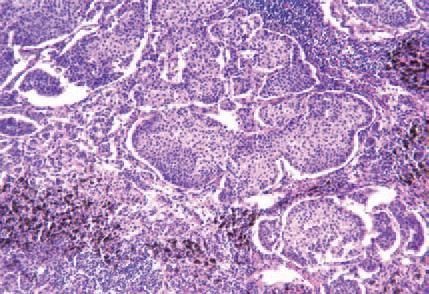
Figure 2 – Right upper lobe wedge resection revealed multiple solid nests bathed by a prominent capillary network. Immunohistochemical stain was positive for synaptophysin and chromogranin.
On follow-up 6 months after surgery, the patient had recovered and was asymptomatic. Despite no significant change in body mass index (21.7 kg/m2 and 21 kg/m2 preoperative and 6 months postoperative, respectively), the patient's lung volumes were significantly increased: FVC was 4 L (70% of predicted) and TLC was 6.8 L (93% of predicted), without significant change in DLCO. MIP was 97 cm H2O (76% of predicted), and MEP was 85 cm H2O (36% of predicted).
A repeated thoracic spine radiograph showed persistent diffuse osteopenia and compression deformity of T4, T6, T8, T10, and T11. The ACTH level had decreased from 143 to 40 pg/mL, and the IGF-I level had increased from 62 to 210 ng/mL (Figure 3). As the patient's levels of ACTH and IGF-I normalized, the TLC increased significantly (Figure 4).
,
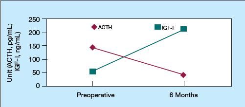
Figure 3 – The decline in ACTH level after surgery was accompanied by an increase in insulin like growth factor I (IGF-I) level. (ACTH, adrenocorticotropic hormone.)
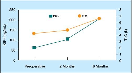
Figure 4 –The increase in insulin like growth factor I (IGF-I) level after surgery was accompanied by an increase in total lung capacity (TLC).
Discussion
We presented this case report at the 2006 annual meeting of the American College of Chest Physicians, and an abbreviated version of this case report was published in Chest.1
Cushing syndrome refers to a condition of hypercortisolism resulting from a primary adrenal disorder or an excess production of ACTH, and it influences the pituitary gland leading to growth hormone deficiency.2 Cushing syndrome can lead to muscular weakness, particularly in the limb muscles.3 Despite severe limb muscle weakness, the respiratory muscles are usually spared in endogenous hypercortisolism.3 Respiratory muscle weakness, however, has been documented in Cushing syndrome resulting from exogenous corticosteroids.4
Our patient presented with restrictive lung disease and weakness of the expiratory muscles. The reduction in lung volume could have been the result of respiratory muscle weakness, obesity, chest wall defects, parenchymal lung disease, or other causes.
After treatment, despite the patient's lower MIP and MEP, his lung volumes increased significantly. The inverse relationship between the respiratory muscle forces and lung volumes delineates 2 important facts. First, the measurement of MIP and MEP is an effort-dependent test, and lower values may be the result of lack of full effort. Second, although expiratory muscle weakness has been documented in similar cases of Cushing syndrome, it has less impact on respiratory function and does not explain the restrictive defect in this situation.
Obesity causes a pattern that is similar to mild restriction. Abdominal compression of the lower lung leads to early airway closure. While TLC and FVC increased in this patient after treatment, his weight did not change, and the abdominal CT scan did not suggest any central obesity.
Chest wall deformities may cause restrictive ventilatory defect, often with a preserved residual volume and normal DLCO. The most common condition is kyphoscoliosis associated with acquired or congenital deformities. The severity of this condition is defined by measurement of the Cobb angle of curvature that is formed by the edges of the vertebral convex.
Our patient had a vertebral compression in the lower thoracic spine, which may have contributed to the ventilatory restriction. The lung volumes, however, started to improve before the vertebroplasty was performed. Furthermore, Cobb angle measurements on lateral chest radiographs before and after vertebroplasty were comparable (Figure 5). Hence, chest wall restriction does not entirely explain the reduced lung volume.
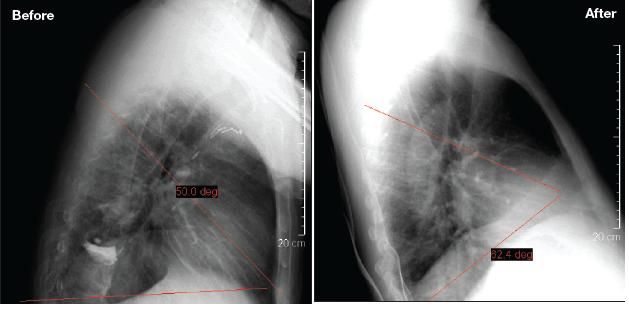
Figure 5 –These lateral chest radiographs were obtained before and after vertebroplasty. The red lines define the Cobb angles. The Cobb angle measurements before and after vertebroplasty were comparable, suggesting that chest wall restriction was not the cause of the patient's reduced lung volume.
Parenchymal lung disorders have a restrictive defect, with reductions in TLC, functional residual capacity (FRC), and residual volume accompanied by reduction in DLCO as a result of mismatching between alveolar ventilation and perfusion. However, the patient's chest CT scan did not suggest any parenchymal lung disease.
Studies have found that adults with growth hormone deficiency diagnosed in childhood had lower FRC and TLC than did matched controls.5 This restrictive defect was related to decreased lung size and not to neuromuscular impairment or chest wall abnormality.6 The corresponding increase in growth hormone level and improvement in lung volumes after surgery raise the possibility that growth hormone causes the improvement in lung volumes in adults.
Several studies of the effect of growth hormone on lung volume have reached conflicting conclusions. While one study found that recombinant growth hormone did not significantly increase lung volumes in patients with growth hormone deficiency,5 other studies found a significant correlation between the growth hormone level and lung mechanics.
For instance, De Troyer and associates6 studied 2 groups of patients with extreme plasma levels of growth hormone-hypopituitarism and acromegaly-and found that growth hormone influences lung volumes and that the loss of growth hormone secretion is responsible for the restrictive ventilatory impairment that is associated with hypopituitarism. Similarly, another study found that the suppression of growth hormone overproduction reduced lung volumes without changing DLCO in patients with acromegaly, compared with matched healthy controls.7
The exact mechanism of the reduced lung volumes in growth hormone deficiency is currently unknown. There is some suggestion that it may be the result of alveolar atrophy.5 Although growth hormone was not found to play a direct role in lung growth in humans, the IGF expression in lung tissues may play an important role in the development of the lungs.8
References:
REFERENCES
1.
Sankri-Tarbichi AG, Saydain G. Reduced lung volumes in a rare case of Cushing syndrome due to thymic carcinoid of the lung.
Chest.
2006; 130:324S-325S.
2.
Tzanela M, Karavitaki N, Stylianidou C, et al. Assessment of GH reserve before and after successful treatment of adult patients with Cushing's syndrome.
Clin Endocrinol (Oxf).
2004;60:309-314.
3.
Ilias I, Torpy DJ, Pacak K, et al. Cushing's syndrome due to ectopic corticotropin secretion:twenty years' experience at the National Institutes of Health.
J Clin Endocrinol Metab.
2005;90:4955-4962.
4.
Mills GH, Kyroussis D, Jenkins P, et al. Respiratory muscle strength in Cushing's syndrome.
Am J Respir Crit Care Med.
1999;160:1762-1765.
5.
Merola B, Longobardi S, Sofia M, et al. Lung volumes and respiratory muscle strength in adult patients with childhood- or adult-onset growth hormone deficiency: effect of 12 months' growth hormone replacement therapy.
Eur J Endocrinol.
1996;135:553-558.
6.
De Troyer A, Desir D, Copinschi G. Regression of lung size in adults with growth hormone deficiency.
Q J Med.
1980;49:329-340.
7.
GarcÃa-RÃo F, Pino JM, DÃez JJ, et al. Reduction of lung distensibility in acromegaly after suppression of growth hormone hypersecretion.
Am J Respir Crit Care Med.
2001;164:852-857.
8.
Stiles AD, D'Ercole AJ. The insulin-like growth factors and the lung.
Am J Respir Cell Mol Biol.
1990;3:93-100.
