- Clinical Technology
- Adult Immunization
- Hepatology
- Pediatric Immunization
- Screening
- Psychiatry
- Allergy
- Women's Health
- Cardiology
- Pediatrics
- Dermatology
- Endocrinology
- Pain Management
- Gastroenterology
- Infectious Disease
- Obesity Medicine
- Rheumatology
- Nephrology
- Neurology
- Pulmonology
Arterial blood gas analysis: A 3-step approach to acid-base disorders
The foundation of arterial blood gas (ABG) analysisconsists of determining whether the patient has acidosis or alkalosis;whether it is a respiratory or metabolic process; and,if respiratory, whether it is a pure respiratory process. If the patient'spH and PCO2 are increased or decreased in the same direction,the process is metabolic; if one is increased while theother is decreased, the process is respiratory. In a number ofclinical situations, pulse oximetry is preferred to ABG analysis.However, pulse oximetry may not be accurate in patients whoare profoundly anemic, hypotensive, or hypothermic. Whilevenous blood gas (VBG) analysis does not provide any informationabout the patient's oxygenation, it can help assessthe level of acidosis or alkalosis. VBG analysis may be particularlyuseful in patients with diabetic or alcoholic ketoacidosis.(J Respir Dis. 2008;29(2):74-82)
ABSTRACT:The foundation of arterial blood gas (ABG) analysis consists of determining whether the patient has acidosis or alkalosis; whether it is a respiratory or metabolic process; and, if respiratory, whether it is a pure respiratory process. If the patient's pH and PCO2 are increased or decreased in the same direction, the process is metabolic; if one is increased while the other is decreased, the process is respiratory. In a number of clinical situations, pulse oximetry is preferred to ABG analysis. However, pulse oximetry may not be accurate in patients who are profoundly anemic, hypotensive, or hypothermic. While venous blood gas (VBG) analysis does not provide any information about the patient's oxygenation, it can help assess the level of acidosis or alkalosis. VBG analysis may be particularly useful in patients with diabetic or alcoholic ketoacidosis. (J Respir Dis. 2008;29(2):74-82)
Correct interpretation of an arterial blood gas (ABG) value can quickly help identify a wide range of illnesses. Most physicians recall learning the Henderson-Hasselbalch equation and how to calculate pH based on the presence of hydrogen ions. However, applying this knowledge in practice on a day-to-day basis can be challenging for even the most learned physician.
In this article, we provide a simple, 3-step approach to evaluate blood gases quickly and correctly, thus allowing valuable information to be rapidly available for evaluating and treating patients.
THE 3-STEP APPROACH
Our 3-step approach can be summarized as follows:
• Step 1: Does the patient have acidosis or alkalosis?
• Step 2: Is the acidosis/alkalosis a respiratory or metabolic process?
• Step 3: If it is respiratory acidosis or alkalosis, is it a pure respiratory process or is there also a metabolic component?
Step 1
If the patient's pH is 7.35 or lower, an acidosis is present. A pH of 7.45 or higher indicates that the patient has an alkalosis.
Step 2
If the PCO2 drives the pH in the opposite direction, there is a primary respiratory process (as PCO2 increases, pH decreases, and vice versa). If the PCO2 and pH move in the same direction, a metabolic process is occurring.<1,2
Step 3
In a pure respiratory process, for every 10 mm Hg change in PCO2, the pH should move in the opposite direction by 0.08 (± 0.02). For example: if the patient's PCO2 is 30 mm Hg (a decrease of 10 mm Hg), the pH should be 7.48 (an increase of 0.08). Or, if the PCO2 is 60 mm Hg (an increase of 20 mm Hg), the pH should be 7.24 (a decrease of 0.16, or 0.08 for each 10 mm Hg rise in PCO2).
If this rule is violated, a second metabolic process is also present. This is often referred to as a mixed process. Step 3 essentially compares the "should be" pH with the actual pH-that is, whether the pH is what it should be based on the rule that for each 10 mm Hg change in PCO2, the pH should change by 0.08. This pH can always be ± 0.02 of the predicted formula's value.
If the patient's pH is not what it should be, is it higher or lower? If it is higher, there must be a concomitant metabolic alkalosis. If the patient's pH is lower, there must be a concomitant metabolic acidosis.
EXAMPLES
Applying this 3-step approach to each case will correctly and quickly identify the underlying acid-base disorder.1,2
Example 1
The patient's pH is 7.56, PCO2 is 20 mm Hg, and PO2 is 110 mm Hg.
•Step 1: The patient's pH is greater than 7.45, indicating that an alkalosis is present.
•Step 2: The PCO2 and pH are moving in opposite directions, so it is a respiratory process.
•Step 3: The PCO2 is decreased by 20 mm Hg, and the pH should be elevated by 2 × 0.08, or 0.16 (± 0.02). Therefore, this is a pure respiratory alkalosis.
•Conclusion: The patient has a simple respiratory alkalosis.
Example 2
The patient's pH is 7.16, PCO2 is 70 mm Hg, and PO2 is 80 mm Hg.
•Step 1: The pH is less than 7.35, so an acidosis is present.
•Step 2: The values are moving in opposite directions, so it is a respiratory process.
•Step 3: Since the PCO2 is increased by 30 mm Hg, the pH should be decreased by 3 × 0.08, or 0.24 (± 0.02), to 7.16, which it has.
• Conclusion: The patient may have a pure respiratory acidosis.
Example 3
The patient's pH is 7.50, PCO2 is 50 mm Hg, and PO2 is 100 mm Hg.
•Step 1: Since the pH is greater than 7.45, an alkalosis is present.
•Step 2: The pH and the PCO2 are moving in the same direction, so this is a metabolic process.
•Step 3: Does not apply, since this is not a respiratory process.
•Conclusion: The patient has a metabolic alkalosis.
Example 4
The patient's pH is 7.25, PCO2 is 25 mm Hg, and PO2 is 90 mm Hg.
•Step 1: The patient has a low pH, so an acidosis is present.
•Step 2: The pH and PCO2 are both decreased. The PCO2 has not driven the pH in the opposite direction, so this process is a metabolic process.
• Step 3: Does not apply, since this is not a respiratory process.
• Conclusion: The patient has a metabolic acidosis.
Example 5
The patient's pH is 7.50, the PCO2 is 20 mm Hg, and the PO2 is 75 mm Hg.
•Step 1: The pH is greater than 7.45, so an alkalosis is present.
•Step 2: The pH and PCO2 are moving in opposite directions, so this is a respiratory process.
•Step 3: The PCO2 has decreased by 20 mm Hg, so the pH should increase by 2 × 0.08, or 0.16, to equal 7.56, but the pH is lower (7.50) than it should be. Therefore, a secondary process, which must be metabolic, is making the patient more acidotic. (Note: This process cannot be respiratory, since we have already determined what the pH should be based on the fall in PCO2.)
•Conclusion: The patient has a primary respiratory alkalosis and a metabolic acidosis. We cannot determine whether the metabolic acidosis is a primary or secondary process, although it often is a compensatory process.
Example 6
The patient's pH is 6.80, PCO2 is 60 mm Hg, and PO2 is 45 mm Hg
•Step 1: Since the pH is less than 7.45, an acidosis is present.
•Step 2: The pH and PCO2 are moving in opposite directions, so this is a respiratory process.
•Step 3: The PCO2 has increased by 20 mm Hg, so the pH should decrease by 2 × 0.08, or 0.16, to equal 7.24. Since the pH is much lower (6.80) than this, there must also be a metabolic acidosis driving the pH down lower than what would be caused by a rise in the PCO2 to 60 mm Hg.
•Conclusion: The patient has a primary respiratory acidosis and a primary metabolic acidosis.
ADDITIONAL CONSIDERATIONS
Both an ABG and a basic metabolic profile are usually needed to completely evaluate a patient's acid-base status and to determine the cause of the metabolic disturbance.
Respiratory acidosis
In a patient with acidosis, determine whether the PCO2 is elevated (respiratory acidosis) or is not as low as would be expected (incomplete compensation for a primary metabolic acidosis). Respiratory acidosis is usually the result of one of the following: airway obstruction, airway spasm, chronic obstructive pulmonary disease (COPD), asthma, massive pulmonary edema, pneumothorax, lung mass, pneumonia, aspiration, oversedation, narcotic overdose, CNS tumor, or neuromuscular disorder.1,2
Remember that eventually there will be compensatory bicarbonate (HCO3) retention in patients who have respiratory acidosis. The kidneys will begin to retain HCO3 within 12 to 16 hours. Maximum compensation, however, takes at least 2 to 3 days.3 HCO3 compensation in respiratory acidosis is as follows:
• Acute: A 1 mEq/L rise in HCO3 for each 10 mm Hg rise in PCO2.
• Chronic: A 3 to 4 mEq/L rise in HCO3 for each 10 mm Hg rise in PCO2.
If the HCO3 value is too high (greater than 30 mEq/L) in a patient with respiratory acidosis, consider the possibility of a chronic respiratory acidosis (COPD, muscular dystrophy, Guillain-Barr syndrome) or a coexistent primary metabolic alkalosis (vomiting, dehydration, nasogastric suctioning).
Respiratory alkalosis
A respiratory alkalosis is the only acid-base disturbance that can possibly have full compensation, thus it is the only acid-base disorder in which a patient's pH can return to normal.1
•Acute: pH rises by 0.08 for every 10 mm Hg fall in PCO2; HCO3 falls by 2 mEq/L for every 10 mm Hg fall in PCO2.
• Chronic: pH begins to approach normal value; HCO3 falls by 5 mEq/ L for every 10 mm Hg fall in PCO2.
Consider the following causes in patients with respiratory alkalosis: anxiety (always a diagnosis of exclusion), aspirin overdose, sepsis, fever, pulmonary embolism, and hypoxia. Other potential causes include hyperthyroidism, pregnancy, liver disease, CNS tumor, pain, and alcohol or drug withdrawal.3,4
Metabolic acidosis
This is usually divided into 2 types. A wide gap acidosis is caused by unmeasured anions and can be best remembered by the mnemonic MUDPILES: methanol, uremia, diabetic ketoacidosis and alcoholic ketoacidosis, paraldehyde, isoniazid and iron, lactic acidosis, ethylene glycol, and salicylates and solvents.
Normal gap acidosis does not have unmeasured anions and, thus, no elevated anion gap. This is because as HCO3 falls, the serum chloride level rises in response, thus the term "hyperchloremic metabolic acidosis." The causes of this type of metabolic acidosis are best remembered by the mnemonic HARDUP: hyperventilation; acid infusion, carbonic anhydrase inhibitors, and Addison disease; renal tubular acidosis; diarrhea; ureterosigmoidostomy (and ileal diversions); and pancreatic fistula or drainage.
Metabolic alkalosis
This kind of alkalosis is usually divided into 2 major types: chlorideresponsive and chloride-unresponsive. A chloride-responsive metabolic alkalosis is corrected by the infusion or oral ingestion of sodium chloride and volume. The most common causes include vomiting, dehydration, HCO3 administration, and diuretic use.
A chloride-unresponsive metabolic alkalosis usually has an underlying endocrinological cause and will not be cured by volume and solute alone. The most commonly encountered causes are Cushing syndrome, corticosteroid use, and hyperaldosteronism.
Mixed processes
Although there are 4 primary acidbase disturbances (2 metabolic and 2 respiratory), there are many more mixed types of disorders if each type and subtype of acidosis and alkalosis is considered. However, only a few mixed acid-base disturbances are commonly seen.
• Primary wide gap metabolic acidosis plus primary respiratory alkalosis: Aspirin overdose and sepsis should always be immediately considered when a wide gap acidosis (resulting from salicylic acid or endotoxininduced lactic acidosis) is seen in association with a primary respiratory alkalosis. Once these 2 potentially fatal conditions have been ruled out, it is more likely that the patient has hypotension (lactic acidosis) and pain (hyperventilation) or is an alcoholic (alcoholic ketoacidosis) and is in withdrawal (hyperventilation).3
• Wide gap metabolic acidosis plus respiratory acidosis: This combination is usually the result of lactic acidosis and hypoventilation and is seen in patients in cardiac arrest who have inadequate or no cardiopulmonary resuscitation and in patients with hypotension who also have pulmonary edema.
• Metabolic acidosis (wide or normal gap) plus metabolic alkalosis: This entity is most commonly seen when a patient has protracted vomiting combined with an underlying meta-bolic acidosis. A vomiting patient with diabetic ketoacidosis would have a metabolic alkalosis and a wide gap acidosis, while a patient with gastroenteritis associated with severe diarrhea and vomiting would have a normal gap acidosis with metabolic alkalosis.
• Primary respiratory alkalosis with a compensatory metabolic acidosis: This is a common compensatory mixed process, other than the hyperventilation seen in a patient who is acidotic. Patients who hyperventilate for a prolonged time begin to compensate within 12 to 24 hours by losing increased quantities of HCO3 in their urine. Thus, a secondary, normal gap acidosis develops in response to a chronic respiratory alkalosis. This mixed process may be seen in patients who are pregnant or who have cirrhosis or anxiety. This is never a wide gap acidosis.
Oxygen saturation
Pulse oximetry is often used to determine a patient's oxygen saturation and to approximate the PO2. It is important for any physician using pulse oximetry to be familiar with some basic principles of this technology. It is also important to know pulse oximetry's limitations and potential problems in special clinical situations.
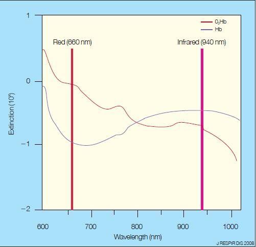
The pulse oximeter generates pulsations of light that are transmitted through tissue (usually a finger, toe, or earlobe) at 400 Hz. Two wavelengths of light are measured at 660 and 940 nm, and the absorption of light in each is determined ( Figure 1).5 Deoxygenated (unsaturated) blood absorbs more red light at 660 nm than does oxygenated (saturated) blood. Oxygenated blood absorbs more UV light at 940 nm than does deoxygenated blood. Both arterial and venous saturations are obtained, and the pulsatile (arterial) and nonpulsatile (venous) absorptions are compared for each wavelength. The resulting measurements are then translated into the oxygen saturation value.5,6

Figure 1 – The hemoglobin (Hb) extinction curve of oxyhemoglobin (O2Hb) with reduced Hb is shown here. Deoxygenated (unsaturated) blood absorbs more red light at 660 nm than does oxygenated (saturated) blood. Oxygenated blood absorbs more UV light at 940 nm than does deoxygenated blood.
The standard settings to calibrate pulse oximeters are based on measurements taken from young, healthy adults with normal hemoglobin levels. Settings are based on data taken when these healthy volunteers had their hemoglobins desaturated from 100% to 80% under close observation. Any saturation below 80% had to be done by extrapolation, since it would have been unethical to induce this level of hypoxemia in humans. When a patient's oxygen saturation is greater than 80%, the pulse oximeters are very accurate and read to within ± 2%. Thus, machines are accurate down to a PO2 of about 50 mm Hg (80% saturation).5,6
Pulse oximeters provide information about the percentage of hemoglobin saturated by oxygen in arterial blood, not the partial pressure of oxygen (which is measured via the ABG). Thus, oxygen saturation measured via pulse oximeter probe is, theoretically, more accurate than oxygen saturation determined by an ABG analysis. This is because pulse oximetry directly measures saturation of almost all of the blood. In contrast, ABG analyses use a calculated saturation based on only 1% to 2% of the blood-the small fraction of oxygen that is dissolved in plasma Many clinicians are not aware of this fact. Rather than truly measuring the saturation of hemoglobin, an ABG analysis provides an estimate.
However, there are several limitations to pulse oximetry. First, any movement by the patient may decrease the oximeter's ability to sense the different wavelengths of light accurately. Second, any device that emits electromagnetic interference, such as MRI equipment, may disrupt the microprocessor in the pulse oximeter probe. Finally, this is a pulse-dependent method of measuring the oxygen saturation; if the patient is hypotensive or severely vasoconstricted, only a weak pulse is present, and the oximeter may not be able to measure oxygen saturation accurately.5,6
ABG analysis versus pulse oximetry
In ABG analysis, oxygen saturation is a calculated value derived by extrapolating from the PO2, which is the amount of dissolved oxygen in blood (only about 1.5% of blood total oxygen content) rather than by directly measuring the oxygen saturation of hemoglobin. It is usually very accurate, but it can provide falsely elevated values in some circumstances, including carbon monoxide poisoning. The comparison between ABG analysis and pulse oximetry in specific clinical settings is described below.
• Healthy patients: In healthy patients, pulse oximetry may estimate true oxygen saturation within 1% to 2%. Thus, pulse oximetry is preferred to ABG analysis because it is highly accurate and noninvasive.7
• Anemia: Even if a patient is anemic, pulse oximetry may be used if the hematocrit is not less than 15%. Once it falls below this value, the pulse oximeter is no longer accurate. In profoundly anemic patients-those with a hematocrit of less than 15% to 20%-ABG analysis is required to accurately measure saturation.5
• Sickle cell anemia: Patients with sickle cell anemia generally have pulse oximetry readings that are reliable within 2% to 4%; thus, pulse oximetry is preferred to ABG analysis in this setting.8
• Hypotension: Hypotension may cause falsely low oxygen saturation readings because of poor arterial pulsation and vasoconstriction. If a patient is in shock, a low pulse oximetry reading does not conclusively indicate hypoxemia; however, a normal saturation reading does confirm good oxygenation.5,7
• Hypothermia: When a patient is hypothermic, cardiac output and peripheral blood flow are decreased, producing an unreliable oxygen saturation reading. A low pulse oximetry reading does not necessarily equate with hypoxemia and should not dictate clinical care.5,7
• Carbon monoxide poisoning: This problem requires special consideration. Neither an ABG analysis, with its customary extrapolated oxygen saturation, nor pulse oximetry provides a true oxygen saturation reading. Carbon monoxide is maximally absorbed at 1 of the 2 wavelengths used to measure oxyhemoglobin (660 nm). An oxygen saturation determined by pulse oximetry is the percentage of hemoglobin saturated by either oxygen or carbon monoxide. Therefore, carbon monoxide produces a falsely elevated oxygen saturation.
If only a pulse oximeter is used, carbon monoxide poisoning will be missed and the patient will appear to be well saturated (with just oxygen). 9 A carbon monoxide level of 50% produces an oxygen saturation of 94%, and saturations above 96% to 97% are seen in more than two thirds of patients. Similarly, a routine ABG analysis does not reflect the amount of hemoglobin saturated with carbon monoxide, because it will merely calculate oxygen saturation by extrapolating from the amount of oxygen dissolved in the blood.
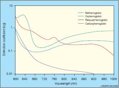
In patients with suspected carbon monoxide poisoning, a cooximeter, which directly measures at least 4 wavelengths instead of 2, is required. A co-oximeter directly measures specific absorption patterns for oxygenated blood, deoxygenated blood, carbon monoxide, and methemoglobinemia (Figure 2).5 The co-oximeter separates out the carbon monoxide and gives an accurate measurement.9 Until recently, co-oximeters were available only in blood gas laboratories; however, highly accurate bedside and portable co-oximeters are now available for emergency department, ICU, and in-the-field use.10

Figure 2 – The hemoglobin extinction curve demonstrates relative absorptions of the 4 major classes of hemoglobin.
• Methemoglobinemia: Methemoglobin absorbs at 660 and 940 nm. As methemoglobin levels increase, the oxygen saturation reading falls toward 85%, where it will stay regardless of the true oxygen saturation. When methemoglobin levels are less than 20 mm Hg, oxygen saturation decreases by 50% of the methemoglobin level. Pulse co-oximetry is now available on portable or bedside monitors, which can readily measure carbon monoxide and methemoglobinemia.5,10
• Cyanide poisoning: This poses another problem. Cyanide does not affect saturation of hemoglobin but does block desaturation. Thus, high oxygen levels and the resultant 100% saturation readings are accurate but do not reflect tissue oxygen utilization. The blood's hemoglobin is completely saturated, but no oxygen is being "unloaded" to the hypoxic tissue, since cellular respiration via the cytochromic-oxidase system is poisoned.11
• Asthma: Many persons who have asthma receive oxygen via their aerosol treatments. Since oxygen saturation levels in the high 90s reflect oxygenation but not ventilation, carbon dioxide retention may be unrecognized. Remember that as patients with an acute exacerbation of asthma tire, they will have increased carbon dioxide levels, but because they are receiving supplemental oxygen, their oxygen saturation will remain high. Initial blood gases will reveal a respiratory alkalosis (such as a pH of 7.5 and a PCO2 of 30 mm Hg). As respiratory failure ensues, carbon dioxide is retained and blood gases normalize (pH of 7.4 and PCO2 of 40 mm Hg) for a short time. However, as the patient tires, the carbon dioxide rises further and produces a respiratory acidosis and, ultimately, respiratory arrest unless emergent interventions are begun.
Be aware that most patients with asthma who are receiving supplemental oxygen have high PO2 and oxygen saturations-until they stop breathing. The treatment of patients with asthma should generally be based on clinical evaluation and spirometry. ABG analysis should only be used when you are unsure of the patient's respiratory status and suspect carbon dioxide retention.
Do not wait for the results of ABG analysis to validate the need to intubate a patient; rely on your clinical judgment, taking into account factors such as the patient's mental status, work of breathing, and spirometric measures.12 Finally, carefully consider whether an ABG analysis is needed, as opposed to just a venous pH and/or a pulse oximetry reading.
VBG versus ABG
When should a venous blood gas (VBG) analysis be used instead of an ABG analysis? In general, in a nonhypotensive patient, the venous pH is equal to the arterial pH minus 0.02 to 0.03. Keep in mind that a VBG analysis does not provide any information about the patient's oxygenation or ventilation, but it can be useful in assessing the level of acidosis or alkalosis. As patients get sicker and more acidotic, the difference between ABGs and VBGs widens, and the venous pH increases to 0.03 to 0.05.
Although a venous pH is still useful for predicting a patient's arterial pH, a venous pH of 7.25 can be seen in a patient with an arterial pH as high as 7.30. If, however, a patient is hypotensive or in shock, there will be significant differences between his or her ABGs and VBGs.13
As patients become hypotensive, their arterial and venous systems become increasingly divergent in pH values. Rising venous carbon dioxide levels disallow the use of a VBG analysis to estimate a hypotensive patient's pH. This arterialvenous difference is maximal during cardiac arrest, when the hypercarbic venous system may create pH differences of up to 0.3. Thus, a venous pH of 7.15 might be seen, while the well-ventilated arterial pH might approximate 7.45.
As a general rule of thumb, a VBG analysis is an excellent choice in patients who are well perfused but whose acid-base status is in question. Clinical examples in which a venous pH is very useful include patients presenting with diabetic ketoacidosis or alcoholic ketoacidosis. A venous sample is also the most appropriate choice in patients for whom a carbon monoxide level is required. Carbon monoxide's half-life is so long (approximately 90 minutes on 100% oxygen) that the venous and arterial levels are equal.13,14
An ABG analysis is required, however, for patients who are in shock, respiratory distress, or cardiac arrest. An ABG analysis in these patients provides accurate pH values and allows determination of ventilation and oxygenation status.
References:
REFERENCES
1.
Narins RG, Emmett M. Simple and mixed acid-base disorders: a practical approach.
Medicine (Baltimore).
1980;59:161-187.
2.
Gluck SL. Acid-base.
Lancet.
1998;352:474-479.
3.
Adrogué HJ, Madias NE. Management of life-threatening acid-base disorders. First of two parts.
N Engl J Med.
1998;338:26-34.
4.
Adrogué HJ, Madias NE. Management of life-threatening acid-base disorders. Second of two parts.
N Engl J Med.
1998; 338:107-111.
5.
McMorrow RC, Mythen MG. Pulse oximetry.
Curr Opin Crit Care.
2006;12:269-271.
6.
Andrews F, Nolan JP. Critical care in the emergency department: monitoring the critically ill patient.
Emerg Med J.
2006;23:561-564.
7.
Lee WW, Mayberry K, Crapo R, Jensen RL. The accuracy of pulse oximetry in the emergency department.
Am J Emerg Med.
2000;18:427-431.
8.
Ortiz FO, Aldrich TK, Nagel RL, Benjamin LJ. Accuracy of pulse oximetry in sickle cell disease.
Am J Respir Crit Care Med.
1999;159:447-451.
9.
Kao LW, Nañagas KA. Carbon monoxide poisoning.
Med Clin North Am.
2005;89:1161-1194.
10.
Barker SJ, Curry J, Redford D, Morgan S. Measurment of carboxyhemoglobin and methemoglobin by pulse oximetry: a human volunteer study.
Anesthesiology.
2006;105:892-897.
11.
Morocco AP. Cyanides.
Crit Care Clin.
2005;21:691-705.
12.
Rodrigo GJ, Rodrigo C, Hall JB. Acute asthma in adults: a review.
Chest.
2004;125:1081-1102.
13.
Gennis PR, Skovron ML, Aronson ST, Gallagher EJ. The usefulness of peripheral venous blood in estimating acid-base status in acutely ill patients.
Ann Emerg Med.
1985;14:845-849.
14.
Brandenburg MA, Dire DJ. Comparison of arterial and venous blood gas values in the initial emergency department evaluation of patients with diabetic ketoacidosis.
Ann Emerg Med.
1998;31: 459-465.
Obesity Linked to Faster Alzheimer Disease Progression in Longitudinal Blood Biomarker Analysis
December 2nd 2025Biomarker trajectories over 5 years in study participants with AD show steeper rises in pTau217, NfL, and amyloid burden among those with obesity, highlighting risk factor relevance.
