- Clinical Technology
- Adult Immunization
- Hepatology
- Pediatric Immunization
- Screening
- Psychiatry
- Allergy
- Women's Health
- Cardiology
- Pediatrics
- Dermatology
- Endocrinology
- Pain Management
- Gastroenterology
- Infectious Disease
- Obesity Medicine
- Rheumatology
- Nephrology
- Neurology
- Pulmonology
Resolution of Papular Mucinosis in a Person With HIV Infection
Lichen myxedematosus, or papular mucinosis, is a skin disorder characterized by accumulation of mucin in the dermis. We present the case of a patient with HIV infection and lichen myxedematosus whose skin condition completely resolved with antiretroviral therapy. [AIDS Reader. 2007;17:418-420]
Lichen myxedematosus, or papular mucinosis, is a skin disorder characterized by accumulation of mucin in the dermis. Lichen myxedematosus has been divided into 3 clinical forms: localized, disseminated, and generalized. Patients with localized lichen myxedematosus present with papular eruptions with a dermal deposition of mucin in a spotty fashion. Lichen myxedematosus has been associated with HIV infection.1-4 We present the case of a patient with HIV infection and lichen myxedematosus whose skin condition completely resolved with antiretroviral therapy.
CASE SUMMARY
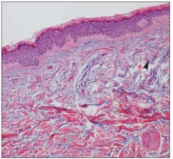
A 45-year-old bisexual man presented with papular lesions on his chest and abdomen in February 2003. The lesions progressed, and in March 2003, a biopsy of the skin confirmed the diagnosis of lichen myxedematosus (Figure 1). In April 2003, oral herpes simplex and diarrhea developed. The result of subsequent testing for HIV antibody the same month was positive.

Figure 1. Biopsy from upper back in March 2003 showing focal mucinosis revealed by an increase of mucin deposits in dermis (arrow). (Stain and original magnification unknown.)
In June 2003, the patient was evaluated for initiating antiretroviral treatment. He had multiple small papular lesions on his chest and abdomen, and mostly on his back. Physical examination showed obesity and cervical lymphadenopathy, but there were no other abnormal findings. Laboratory findings revealed a white blood cell count of 3500/µL; hematocrit, 38.3%; platelet count, 105,000/µL; CD4+ cell count, 58/µL; and HIV RNA level, 100,000 copies/mL.
He began a twice-daily antiretroviral regimen comprising the lopinavir/ritonavir coformulation 400/100 mg plus a zidovudine/ lamivudine fixed-dose combination 300/100 mg. In September 2003, the twice-daily zidovudine/lamivudine fixed-dose combination was replaced by a once-daily fixed-dose combination of tenofovir/emtricitabine 300/200 mg.
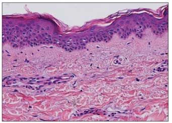
By late October 2003, 4 months after the start of antiretroviral therapy, the patient’s skin lesions had markedly diminished. In January 2004-after 7 months of antiretroviral therapy-the lesions has resolved completely. The results of a second skin biopsy in February 2004 showed no evidence of lichen myxedema tosus (Figure 2). The patient’s skin remains lesion-free to date, as of May 2007.

Figure 2. Biopsy from upper back in February 2004 does not show evidence of mucin or increase in the number of fibroblasts. (Stain and original magnification unknown.)
DISCUSSION
A PubMed search of the literature for published cases of lichen myxedematosus associated with HIV infection found reports of 16 cases. These cases, including ours reported here, are summarized in the Table. In these 17 cases, most patients were homosexual men (15 of 17); CD4+ cell counts of less than 100/µL were noted in 11 (69%) of the 16 whose CD4 count was known at the time of diagnosis of lichen myxedematosus. Two patients treated with zidovudine monotherapy experienced regression of their lesions over a 4-year period.2,3 Our patient, who received a more powerful antiretroviral regimen, had complete resolution of the lichen myxedematosus in 7 months. In the reported cases, homosexual sex and a CD4+ cell count below 100/µL stand out as unique characteristics.
CONCLUSION
The cause of lichen myxedematosus in persons with HIV infection remains unknown. Since 1992, when Ruiz-Rodriguez and colleagues4 reported the first case of lichen myxedematosus in an HIV-infected person, the options for the effective treatment of HIV infection have increased dramatically. The association of lichen myxedematosus with HIV infection warrants consideration of performing a skin biopsy to confirm the diagnosis in HIV-infected patients with papular lesions. Our experience with complete resolution in 7 months after highly active antiretroviral therapy suggests that lichen myxedematosus may be immunologically mediated.
No potential conflict of interest relevant to this article was reported by the authors.
References:
References1. Biro DE, Lynfield YC, Heilman ER. Papular mucinosis and human immunodeficiency virus infection. Cutis. 1995;55:113-114.
2. DomÃnguez Auñon JD, Postigo Llorente C, Llamas Martin R, et al. Lichen myxoedematosus associated with human immunodeficiency virus infection-report of two cases and review of the literature. Clin Exp Dermatol. 1997;22:265-268.
3. Rongioletti F, Ghigliotti G, Marchi R, Rebora A. Cutaneous mucinoses and HIV infection. Br J Dermatol. 1998;139:1077-1080.
4. Ruiz-Rodriguez R, Maurer T, Berger T. Papular mucinosis and human immunodeficiency virus infection. Arch Dermatol. 1992;128:995-996.
5. Tarantini G, Zerboni R, Muratori S, et al. Lichen myxoedamatosus in a patient with AIDS. Br J Dermatol. 1996;134:1122-1124.
6. Moragon M, Sevila A, Colomina J, et al. Papular mucinosis in HIV-infected patients. AIDS. 1995;9:535-536.
Â
