- Clinical Technology
- Adult Immunization
- Hepatology
- Pediatric Immunization
- Screening
- Psychiatry
- Allergy
- Women's Health
- Cardiology
- Pediatrics
- Dermatology
- Endocrinology
- Pain Management
- Gastroenterology
- Infectious Disease
- Obesity Medicine
- Rheumatology
- Nephrology
- Neurology
- Pulmonology
Cryptosporidiosis: Still a Problem
A 22-year-old man with a history of AIDS, gastritis, and perianal warts presented to his primary care physician with a complaint of worsening dyspepsia and diarrhea.
A 22-year-old man with a history of AIDS, gastritis, and perianal warts presented to his primary care physician with a complaint of worsening dyspepsia and diarrhea. The patient denied fever, nausea, vomiting, bloody stools, melena, weight loss, and change in appetite. He had a history of poor adherence to medication and had stopped his antiretroviral regimen of zidovudine, lamivudine, and nelfinavir several weeks before.
Findings from the patient’s examination were notable for normal vital signs and mild abdominal tenderness on palpation. No guarding or rebound was present. Laboratory testing revealed a white blood cell count of 2850/µL; normal hemoglobin level and platelet count; serum protein, 8.7 g/dL (normal, 5.5 to 8.0); serum bilirubin, 1.5 g/dL (normal, 0.2 to 1.3); serum aspartate aminotransferase, 57 g/dL (normal, 0 to 45); and serum alanine aminotransferase, 69 g/dL (normal, 0 to 45). Results of tests for antibodies to hepatitis A, B, and C viruses were negative, as were tests for hepatitis B surface antigen. His CD4+ cell count was 35/µL with a CD4 percentage of 4%, and his HIV RNA level was 544,590 copies/mL. Stool culture and test results for Clostridium difficile toxin were negative. A stool test for ova and parasites showed “few Giardia lamblia cysts and trophozoites” and “moderate Cryptosporidium species.” Fecal smears for Cyclospora and microsporidia were negative.
The patient was given prescriptions for loperamide, metronidazole, and paromomycin for treatment of giardiasis and cryptosporidiosis. He was also advised to resume his antiretroviral therapy.
Over the next several weeks, he was intermittently adherent to his antiparasitic drugs, his antiretrovirals, and follow-up appointments. Subsequent stool studies for ova and parasites were negative for Giardia, but smears for Cryptosporidium remained positive. His diarrhea worsened, and his weight decreased from 77.4 to 68.1 kg (172 to 151.3 lb). His CD4+ cell count declined to 21/µL, and his HIV RNA level remained high at 295,831 copies/mL. He became progressively weaker, with dizziness on standing, and was admitted to the hospital.
In the hospital, the patient’s stool smear for Cryptosporidium and fecal enzyme immunoassay (EIA) for Cryptosporidium antigen were both positive. He was given paromomycin and azithromycin, and treatment with antiretrovirals was restarted. He also received intravenous fluids and tincture of opium. Fever, nausea, and vomiting developed early in his hospital stay, but all of his symptoms, including his diarrhea, resolved.
The patient was discharged to home but again was poorly adherent to his medications. His CD4+ cell count remained low at 22/µL, and his HIV RNA level remained high at 157,057 copies/mL. Diarrhea and dehydration recurred, necessitating readmission to the hospital. A stool smear for Cryptosporidium was negative this time, but a fecal EIA for Cryptosporidium antigen was still positive. He had initial improvement with the addition of azithromycin and the resumption of paromomycin and his antiretroviral therapy, but his condition then worsened despite increasing doses of tincture of opium. His weight loss continued, and he required total parenteral nutrition.
DISCUSSION
Cryptosporidiosis, primarily a GI disease caused by Cryptosporidium, is one of the most common diarrheal illnesses in the world.1,2 Although the disease is self-limited in immunocompetent persons, it can be a devastating and potentially life-threatening infection in those who are immunodeficient, especially those with HIV infection.3,4 Even though the prevalence of cryptosporidiosis among those living with HIV has been declining since the introduction of highly active antiretroviral therapy,4 it is still a serious problem in persons who have undiagnosed HIV infection, in those who are nonadherent to therapy or have highly resistant virus, and in persons in underdeveloped countries in which access to antiretroviral therapy is limited.
The ParasiteCryptosporidium species are intracellular parasites of the phylum Apicomplexa. The most common of the 16 currently recognized species that cause infection in humans was previously classified as Cryptosporidium parvum. There are 2 genotypes of this organism: genotype 1 infects humans; genotype 2 infects humans, bovines, and other mammalian hosts. The human genotype 1 has been reclassified as Cryptosporidium hominis.5
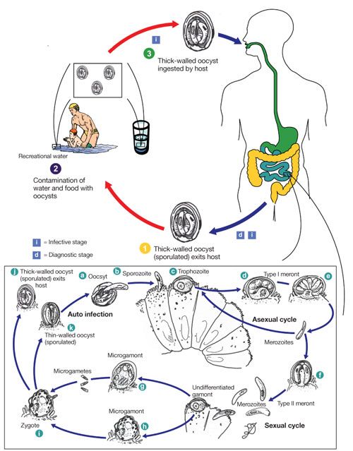
The parasite completes its entire life cycle within the host, beginning with the ingestion of the oocyst forms (Figure 1). The oocysts rupture in the GI tract, releasing sporozoites, which adhere to intestinal epithelial cells and invade them, replicating asexually into merozoites. The merozoites are released into the lumen and either infect other GI epithelial cells or differentiate into gametocytes (gamonts), which reproduce sexually to regenerate oocysts, which can again release sporozoites restarting the cycle. Oocysts are excreted in feces; ingestion of these oocysts by another host spreads the infection.

Figure 1. Life cycle of Cryptosporidium parvum and Cryptosporidium hominis. Cryptosporidium stages were reproduced from Juranek DD. Cryptosporidiosis. In: Strickland GE, ed. Hunter’s Tropical Medicine and Emerging Infectious Diseases. 8th ed. Philadelphia: WB Saunders; 2000. Originally adapted from the life cycle that appears in Current WL, Garcia LS. Cryptosporidiosis. Clin Microbiol Rev. 1991;4:325-358. (From Centers for Disease Control and Prevention, DPDx Parasite Image Library, http://www.dpd.cdc.gov/dpdx.)
EPIDEMIOLOGY
Before the introduction of highly active antiretroviral therapy, 3% to 4% of patients with AIDS in the United States contracted this infection. Rates have declined since antiretroviral therapy was introduced.3
Because the Cryptosporidium oocyst is shed in feces, the infection is spread by person-to-person contact, infected water and food, and animals. Modes of person-to-person contact in the spread of Cryptosporidium include fecal-oral contact as a result of inadequate hygiene, anal-oral sexual contact, and fomites. Infected water sources, both for ingestion and for recreation, have been a major source of community outbreaks of cryptosporidiosis (related to accidental fecal contamination),1 given the hardiness of the oocyst form and the small number of organisms needed to establish infection. Washing of food (eg, raw produce) in contaminated water can also spread the infection. The oocysts can remain infectious for up to 6 months if kept moist6 and are unaffected by the concentrations of chlorine in treated tap water7 or recreational water sources.8 Measures such as filtering, ultraviolet radiation, and ozone treatment are necessary to remove or kill the parasites.9PATHOGENESIS
The precise mechanisms by which Cryptosporidium causes disease in humans are not well understood. However, both impaired intestinal absorption and enhanced secretion are seen in cases of cryptosporidial diarrhea.3 The increased severity of disease in persons with AIDS is likely related to the importance of cell-mediated immunity in both the response and the development of immunity to cryptosporidial infection. A key part of the immune response to the parasite is the role of CD4+ T lymphocytes.10 This is evidenced by the inverse correlation between the number of CD4+ T lymphocytes and the severity of cryptosporidiosis, with more serious symptoms in patients with lower CD4+ cell counts.11 In addition, increases in intestinal mucosal CD4+ cell counts have been seen in AIDS patients with cryptosporidiosis who are on effective antiretroviral therapy regimens and are able to clear the parasite.12CLINICAL FEATURES
The hallmark of cryptosporidiosis is diarrhea, often accompanied by abdominal pain. In some cases, other systemic symptoms, such as fever, malaise, nausea, vomiting, and loss of appetite, are seen. A study of a cohort of patients with HIV infection showed 4 patterns of intestinal disease: asymptomatic infection, transient infection (diarrhea of less than 2 months’ duration, with complete remission of symptoms and clearance of the parasite from feces), chronic diarrhea (diarrhea for more than 2 months, with persistence of the parasite in feces), and fulminant infection (diarrhea of 2 L or more in volume per day).13 The severity of infection varies by the CD4+ cell count, with the most serious manifestations occurring in patients with lower counts.14Cryptosporidium can also cause extraintestinal disease, with biliary infection being the most common. Biliary disease is usually seen in patients with CD4+ cell counts of less than 50/µL and is associated with increased mortality.15 Associated symptoms include right upper quadrant pain, nausea, vomiting, and fever. The alkaline phosphatase level is usually elevated; serum bilirubin and transaminase levels may also be high. Endoscopic retrograde cholecystopancreatography is usually needed for initial diagnosis, which can be confirmed by identifying the organisms in biopsy specimens. Other foci of extraintestinal disease include the lungs,16 middle ear,17 pancreas,18 and stomach.19DIAGNOSIS
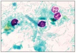
Cryptosporidiosis is usually diagnosed by examination of fecal specimens. The oocyst can be seen with acid-fast staining (Figure 2); a modified Ziehl-Neelsen stain is the one most commonly used.20 The oocysts are recognized by pink or red staining as opposed to the green or blue of the fecal debris. However, because other parasites, such as Isospora and Cyclospora, also have a similar appearance with acid-fast staining, Cryptosporidium must be identified by its smaller oocyst (4 to 6 µm in diameter), which distinguishes it from the other parasites. It must be remembered that the acid-fast staining is not a part of most laboratories’ routine examination of feces for ova and parasites21; it is necessary to order specific tests for Cryptosporidium in most cases.

Figure 2.Oocysts of Cryptosporidium parvum stained by the modified acid-fast method. Against a blue-green background, the oocysts stand out in a bright red stain. Sporozoites are visible inside the two oocysts to the right. (From the Centers for Disease Control and Prevention. DPDx Parasite Image Library, http://www.dpd.cdc.gov/dpdx.)
Sensitivity and specificity are enhanced by use of immunofluorescence assays for Cryptosporidium oocysts or EIA for Cryptosporidium antigen. As noted in the case presented, even the specific microscopic test for the parasite can be negative in the presence of the disease; the use of the immunoassay enhances diagnosis. Polymerase chain reaction–based genotype testing has also been used but is not generally available outside of research laboratories.9TREATMENT
Rehydration with fluids and electrolytes is an important and vital part of care for patients with cryptosporidiosis. In addition, antimotility agents help reduce the diarrhea; in severe disease, agents such as tincture of opium are usually necessary. Octreotide has been shown to be effective in reducing AIDS-related diarrhea; however, because of its high cost and lack of evidence of consistently higher effectiveness than other agents,22 it is generally used only in cases that are refractory to all other agents.
Unfortunately, there is no antiparasitic agent that has been found to be routinely effective for curing this disease. A recent Cochrane review of prevention and treatment of cryptosporidiosis in immunocompromised patients looked at the literature on the use of several agents: paromomycin, nitazoxanide, macrolides, rifabutin/rifamaxin, and bovine immunoglobulin.23 This systemic review of randomized controlled trials found no clear evidence of efficacy among any of these agents in immunocompromised persons. However, given that studies have shown that nitazoxanide does lead to a significant clearance of the parasite in HIV-negative persons, the authors concluded that it is worth considering for use pending further studies in immunocompromised patients. The drug is FDA-approved for use in children with cryptosporidiosis.
Effective antiretroviral therapy has been shown to lead to improvement in diarrhea.24,25 It appears that along with their role in restoration of the immune response, protease inhibitors have anticryptosporidial activity in vitro26; a regimen including one of these drugs should be used whenever possible. If there is a lack of response to antiretrovirals, consideration should be given to the possibility of malabsorption; therapeutic drug levels may be helpful in evaluating this. Also, as in the case presented, adherence is vital to the success of treatment. Until more effective antiparasitic drugs are developed, antiretroviral drugs will remain the mainstay of treatment for cryptosporidiosis in patients with AIDS.
FOLLOW-UP
The case patient was discharged from the hospital to a skilled nursing facility, where he remained for several months. While there, he admitted that he had been surreptitiously disposing of his antiparasitic medications and antiretrovirals while he was in the hospital. However, he had resumed taking them in the skilled nursing facility and took them regularly. His CD4+ cell count rose to 182/µL, and his HIV RNA level decreased to 1053 copies/mL. His stool became negative for Cryptosporidium, and he was discharged to home.
References:
References1. Meinhardt PL, Casemore DP, Miller KB. Epidemiologic aspects of human cryptosporidiosis and the role of waterborne transmission. Epidemiol Rev. 1996;18:118-136.
2. Doganci T, Araz E, Ensari A, et al. Detection of Cryptosporidium parvum infection in childhood using various techniques. Med Sci Monit. 2002;8:MT223-MT226.
3. Chen XM, Keithly JS, Paya CV, LaRusso NF. Cryptosporidiosis. N Engl J Med. 2002;346:1723-1731.
4. Clark DP. New insights into human cryptosporidiosis. Clin Microbiol Rev. 1999;12:554-563.
5. Morgan-Ryan UM, Fall A, Ward LA, et al. Cryptosporidium hominis n sp (Apicomplexa: Cryptosporididae) from Homo sapiens. J Eukaryot Microbiol. 2002;49:433-440.
6. Fayer R, Trout JM, Jenkins MC. Infectivity of Cryptosporidium parvum oocysts stored in water or at environmental temperatures. J Parasitol. 1998;84:1165-1169.
7. Quinn CM, Betts WB. Longer term viability of chlorine-treated Cryptosporidium oocysts in tap water. Biomed Lett. 1993;48:315-318.
8. Carpenter C, Fayer R, Trout J, Beach MJ. Chlorine disinfection of recreational water for Cryptosporidium parvum. Emerg Infect Dis. 1999;5:579-584.
9. Sunnotel O, Lowery CJ, Moore JE, et al. Cryptosporidium. Lett Appl Microbiol. 2006;43:7-16.
10. Riggs MW. Recent advances in cryptosporidiosis: the immune response. Microbes Infect. 2002;4:1067-1080.
11. Flanigan T, Whalen C, Turner J, et al. Cryptosporidium infection and CD4 counts. Ann Intern Med. 1992;116:840-842.
12. Schmidt W, Wahnschaffe U, Schafer M, et al. Rapid increase of mucosal CD4 T cells followed by clearance of intestinal cryptosporidiosis in an AIDS patient receiving highly active antiretroviral therapy. Gastroenterology. 2001;120:984-987.
13. Manabe YC, Clark DP, Moore RD, et al. Cryptosporidiosis in patients with AIDS: correlates of disease and survival. Clin Infect Dis. 1998;27:536-542.
14. Farthing MJ. Clinical aspects of human cryptosÂporiÂdiosis. Contrib Microbiol. 2000;6:50-74.
15. Vakil NB, Schwartz SM, Buggy BP, et al. Biliary cryptosporidiosis in HIV-infected people after the waterborne outbreak of cryptosporidiosis in Milwaukee. N Engl J Med. 1996;334:19-23.
16. Clavel A, Arnal AC, Sanchez EC, et al. Respiratory cryptosporidiosis: case series and review of the literature. Infection. 1996;24:341-346.
17. Dunand VA, Hammer SM, Rossi R, et al. Parasitic sinusitis and otitis in patients infected with human immunodeficiency virus: report of five cases and review. Clin Infect Dis. 1997;25:267-272.
18. Teare JP, Daly CA, Rodgers C, et al. Pancreatic abnormalities and AIDS related sclerosing cholangitis. Genitourin Med. 1997;73:271-273.
19. Rossi P, Rivasi F, Codeluppi M, et al. Gastric involvement in AIDS associated cryptosporidiosis. Gut. 1998;43:476-477.
20. Henriksen SA, Pohlenz JF. Staining of cryptosporidia by a modified Ziehl-Neelsen technique. Acta Vet Scand. 1981;22:594-596.
21. Leav BA, Mackay M, Ward HD. Cryptosporidium species: new insights and old challenges. Clin Infect Dis. 2003;36:903-908.
22. Simon DM, Cello JP, Valenzuela J, et al. Multicenter trial of octreotide in patients with refractory acquired immunodeficiency syndrome-associated diarrhea [published correction appears in Gastroenterology. 1995;109:1024]. Gastroenterology. 1995;108:1753-1760.
23. Abubakar I, Aliyu SH, Arumugam C, et al. Prevention and treatment of cryptosporidiosis in immunocompromised patients. Cochrane Database Syst Rev. 2007;24:CD004932.
24. Carr A, Marriott D, Field A, et al. Treatment of HIV-1-associated microÂsporidiosis and cryptosporidiosis with combination antiretroviral therapy. Lancet. 1998;351:256-261.
25. Maggi P, Larocca AM, Quarto M, et al. Effect of antiretroviral therapy on cryptosporidiosis and microsporidiosis in patients infected with human immunodeficiency virus type I. Eur J Clin Microbiol Infect Dis. 2000;19:213-217.
26. Hommer V, Eichholz J, Petry F. Effect of antiretroviral protease inhibitors alone, and in combination with paromomycin, on the excystation, invasion, and in vitro development of Cryptosporidium parvum [published correction appears in J AntiÂmicrob Chemother. 2003;52:535]. J Antimicrob Chemother. 2003;52:359-364.
