- Clinical Technology
- Adult Immunization
- Hepatology
- Pediatric Immunization
- Screening
- Psychiatry
- Allergy
- Women's Health
- Cardiology
- Pediatrics
- Dermatology
- Endocrinology
- Pain Management
- Gastroenterology
- Infectious Disease
- Obesity Medicine
- Rheumatology
- Nephrology
- Neurology
- Pulmonology
Staphylococcal and Streptococcal Infections
Numerous infections that are generally caused by Staphylococcus aureus also may be caused by Streptococcus species and vice versa.
Key words:Staphylococcus aureus, β-Hemolytic streptococci, Viridans streptococci, Impetigo, Cellulitis, Erysipelas, Bacterial parotiditis, Brodie abscess
Numerous infections that are generally caused by Staphylococcus aureus also may be caused by Streptococcus species and vice versa. The nares are a common reservoir for S aureus, and infection often occurs from hygiene lapses, either by autologous or exogenous exposure to the pathogen. The organism enters through breaks in the skin and begins its infectious process. Streptococci are transmitted from person to person by skin contact and then may colonize the nose and throat.
Impetigo
Impetigo is often caused by S aureus infection, although β-hemolytic streptococci, primarily Streptococcus pyogenes, also can cause it. Impetigo affects all age groups, with the pediatric population being most affected. The bullous form is most common in infants and children younger than 2 years, and the nonbullous form is most common in children aged 2 to 6 years. Health issues, such as declining cardiovascular function, poor circulation, diabetes, obesity, cancer, immunodeficiency, poor circulation, and xerosis can contribute to the development of impetigo and related bacterial infections in older adults (ie, older than 65 years). Group G β-hemolytic streptococci (eg, Streptococcus dysgalactiae subsp equisimilis) are an increasingly common cause of skin and soft tissue infection in this population group.
The rash (nonbullous form) appears as a vesiculopustular eruption with honey-colored crusted erosions that may turn brown as the rash evolves and resolves. Although the diagnosis can be made clinically, it may be helpful to confirm the causative organism, especially in an era in which methicillin-resistant S aureus (MRSA) infection has become so prevalent. Confirmation is done by performing a Gram stain and culture of exudate from the lesions. Treatment typically consists of applying topical mupirocin or fusidic acid to the rash after cleansing the area of crusts and debris using soapy water.1 Oral antibiotics such as cephalexin and clindamycin may be considered in moderate to severe disease, but evidence is lacking on whether oral antibiotics offer more benefit than topical antibiotics even in the setting of extensive disease.1
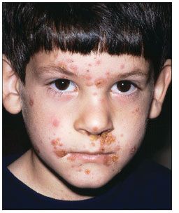
Figure 1 –Impetigo on the face of this 7-year-old boy developed as a complication of chickenpox. (Photo courtesy of Robert P. Blereau, MD.)
Figure 1 shows impetigo secondary to chickenpox on the face of a 7-year-old boy. Figure 2 shows hyperpigmentation and scale on the forehead of an older man.
Cellulitis and erysipelasS aureus and S pyogenes are often implicated in cellulitis and erysipelas. Although all age groups are affected, older persons are at higher risk for these infections. Erythema, pain, swelling, and warmth are common symptoms. Fever-if only low-grade-also may be present. Cellulitis (Figure 3), which typically affects the lower legs, involves the subcutaneous fat and is often less well demarcated than erysipelas, which affects the skin of the lower extremities (Figures 4 and 5) and, less often, the facial area (Figure 6).
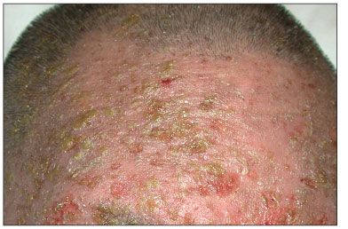
Figure 2 –Scales and hyperpigmentation characteristic of the later phase of impetigo are seen on the forehead of this older man. (Photo courtesy of Noah S. Scheinfeld, MD, JD. Overview adapted from Scheinfeld NS. Consultant. 2007;47:178.)
Cutaneous signs of infection can include orange-peel (peau d’orange) skin and ruddy, erythematous vesicles and/or bullae. Red streaks radiating from an infected area represent progression of the infection into the lymphatic system. The diagnosis is made on the basis of clinical evidence. It is rare that a definitive diagnosis can be made from examination of aspirate or from culturing blood.2
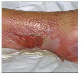
Figure 3 – This example of cellulitis shows inflammation, blistering, and possible lymphatic involvement. (Photo courtesy of Noah S. Scheinfeld, MD, JD.)
Clindamycin, trimethoprim/sulfamethoxazole, and doxycycline are being recommended by the Infectious Diseases Society of America for treatment of cellulitis with purulent drainage or similar skin and soft tissue infections caused by S aureus (assumed to be methicillin-resistant and treated empirically).2 An agent that has activity against β-hemolytic streptococci is recommended for cellulitis in which streptococcal involvement is suspected.2 Nafcillin or cefazolin is recommended for streptococcal cellulitis, and penicillin or a cephalosporin is the first-line treatment for streptococcal erysipelas. Substitutes in persons with penicillin allergy are vancomycin and clindamycin.3
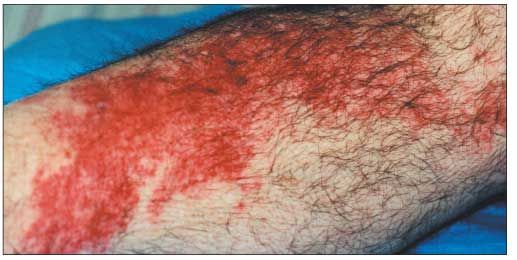
Figure 4 –An erythematous and edematous rash characteristic of streptococcal erysipelas on the lower leg of a 60-year-old man is shown here. (Photo courtesy of Sunita Puri, MD.)
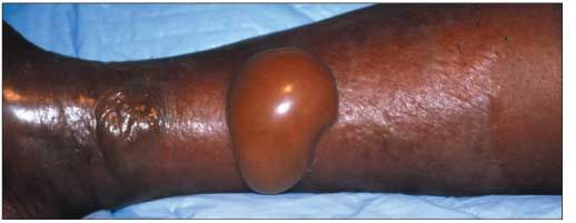
Figure 5 –This example of erysipelas, caused by Staphylococcus aureus, is characterized by bullous lesions. (Photo courtesy of David Effron, MD.)
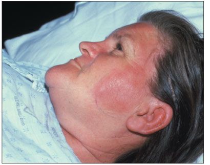
Figure 6 –This example of facial erysipelas shows an indurated and erythematous lesion that might be mistaken for cellulitis, although erysipelas is more demarcated than cellulitis and affects the skin rather than underlying tissue. (Photo courtesy of David Effron, MD.)
Suppurative bacterial parotiditis
Suppurative bacterial parotiditis (Figure 7) commonly occurs in elderly adults who are dehydrated as a consequence of treatment with diuretic or anticholinergic agents or because of surgery. S aureus is most often the cause, followed by viridans streptococci. Xerostomia and procedures that obstruct the Stensen duct contribute to the infectious process by impeding clearance of colonizing bacteria in the oral cavity. Clinical symptoms are intense, unilateral swelling of the parotid gland; erythema; and tenderness. A purulent discharge often exudes from the Stensen duct.
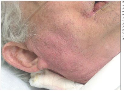
Figure 7 –Erythema and induration extend from the jaw line to the middle of the neck in this example of acute bacterial parotiditis in an elderly man. (Photo courtesy of Andrew Koon, MD, Andrew Bagg, MD, Kevin O’Brien, MD, and Carrie Vey, MD. Overview adapted from Photoclinic in Consultant. 2006;46:1405-1406.)
Empiric therapy that is active against S aureus is first-line treatment, although secretions from the gland should be obtained and cultured to rule out infection by other organisms, including streptococci, gram-negative and anaerobic organisms, and MRSA. Interventions, such as gland massage, may be needed to facilitate drainage of an infected Stensen duct. A CT scan or an ultrasonogram may be required to confirm diagnosis (Figure 8), especially in patients who are unresponsive to therapy within the first 72 hours. Depending on the results of imaging studies-such as identification of complicated infection-surgical intervention may be required.
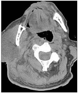
Figure 8 –A CT scan of the head of the patient shown in Figure 7 reveals diffuse swelling and inflammation of the right parotid gland. The inflammation is seen infiltrating the surrounding muscle and fat planes and extending toward the region of the carotid sheath. (Photo courtesy of Andrew Koon, MD, Andrew Bagg, MD, Kevin O’Brien, MD, and Carrie Vey, MD. Overview adapted from Photoclinic in Consultant. 2006;46:1405-1406.)
Brodie abscess
In the pediatric population, S aureus is responsible for 90% of cases of Brodie abscess (Figure 9), although in neonates and infants, group B streptococci also are associated with this infectious process. Boys are more often affected than girls, and the most common sites of involvement are the metaphysis of the distal and proximal tibia, distal femur, and the distal and proximal fibula. The primary differential diagnosis is osteoid osteoma. A distinguishing feature of Brodie abscess is that the radiolucent nidus is larger than that of osteoid osteoma, which may contain a centrum of calcification.
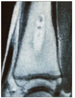
Figure 9 –A T2-weighted inversion recovery MRI scan shows a well-marginated intramedullary lesion indicative of Brodie abscess in a 15-year-old boy. Extensive edema of the tibial metaphysis and early periostitis are evident. (Photo courtesy of Joel M. Schwartz, MD. Overview adapted from Photoclinic in Consultant. 1998;38:2560-2561.)
The abscess develops as a result of immunological containment of acute hematogenous osteomyelitis. The infection is walled off and, thus, a well-corticated margin can be seen on a radiograph. Hematogenous dissemination of infection is more common in pediatric patients than in adult patients because the extremities are more vascular; in adults, bone marrow is less vascular and more fatty.
Treatment consists of surgical therapy and antibiotic therapy.
References:
REFERENCES
1. Koning S, Verhagen AP, van Suijlekom-Smit LW, et al. Interventions for impetigo. Cochrane Database Syst Rev. 2004;(2):CD003261.
2. Liu C. IDSA clinical practice guidelines for the treatment of MRSA infections in progress. Presented at: the joint 48th Annual Interscience Conference on Antimicrobial Agents and Chemotherapy/Infectious Diseases Society of America 46th Annual Meeting; October 26-28, 2008; Washington, DC.
3. Stevens DL, Bisno AL, Chambers HF, et al; Infectious Diseases Society of America. Practice guidelines for the diagnosis and management of skin and soft-tissue infections [published corrections appear in Clin Infect Dis. 2005;41:1830 and Clin Infect Dis. 2006;42:1219]. Clin Infect Dis. 2005;41:1373-1406.
