- Clinical Technology
- Adult Immunization
- Hepatology
- Pediatric Immunization
- Screening
- Psychiatry
- Allergy
- Women's Health
- Cardiology
- Pediatrics
- Dermatology
- Endocrinology
- Pain Management
- Gastroenterology
- Infectious Disease
- Obesity Medicine
- Rheumatology
- Nephrology
- Neurology
- Pulmonology
Pneumocystis Pneumonia in a Person With a Low CD4 Count After Stopping Antiretroviral Therapy
This gay white man was your patient several years ago. He originally presented to you in 1995 with diffuse cutaneous Kaposi sarcoma (KS) and a CD4+ cell count of 15/µL.
This gay white man was your patient several years ago. He originally presented to you in 1995 with diffuse cutaneous Kaposi sarcoma (KS) and a CD4+ cell count of 15/µL. HIV infection had been diagnosed 5 years before, but the patient felt well at the time and did not want to "deal with" the medical system. He had no symptoms until purple lesions developed on his torso, face, and legs. He came to your office for the first time saying, "I know I have AIDS, and I'm ready to do something about it."
You prescribed an indinavir-based 3-drug regimen as his initial antiretroviral therapy. You also prescribed trimethoprim/sulfamethoxazole (TMP/SMX) for prophylaxis against Pneumocystis pneumonia (PCP) and azithromycin to protect against Mycobacterium avium complex (MAC) infection. Your patient was adherent: he never missed a dose and did not complain about the number of pills or taking medications on an empty stomach. His HIV RNA level dropped from 634,262 copies/mL to less than 400 copies/mL. His CD4+ cell count rose slowly but steadily from 15/µL to more than 500/µL. His KS lesions completely disappeared. His antiretroviral regimen eventually became more convenient by boosting the indinavir with low-dose ritonavir, and his PCP and MAC prophylaxis was discontinued.
After 9 years in your care, the patient decided to move to another state where he was hoping to get a new job. He took a medical letter and copy of the results of his laboratory work to give to his next doctor.
More than 2 years passed before you saw this patient again. Four months ago, he presented to your emergency department (ED) noting 3 weeks of a dry cough and worsening shortness of breath. He was not sure if he had had a fever at home, but his temperature in the ED was 38.8°C (101.8°F). The patient had a resting oxygenation saturation of 95% on room air, which dropped to 81% when he got up and walked. His respiratory rate was 21 breaths per minute, and his heart rate was 110 beats per minute. He looked thin and reported a 6.8-kg (15-lb) weight loss over the past 4 months. He had no oral thrush, and his lungs were clear to auscultation. There was no evidence of KS. Chest films revealed diffuse interstitial lung disease. He was admitted with a diagnosis of pneumonia, and treatment for PCP was started with TMP/SMX and a tapering course of corticosteroids. He was also treated with antibiotics for community-acquired pneumonia.
At the hospital, he greeted you warmly despite his obvious shortness of breath. He revealed that the job in the other city had not worked out and that he had come back home after only a few months. He had not received medical care during his time away and because he had been feeling so well for so long, he did not call to renew his medications when they ran out. He ate well, exercised, and enjoyed not taking any pills for the first time in many years. "I guess I should have called you," he panted from behind his oxygen mask.
The patient underwent bronchoscopy the following day, and the washings from the bronchoalveolar lavage revealed many organisms of Pneumocystis jirovecii (formerly Pneumocystis carinii).
INCIDENCE AND EPIDEMIOLOGY OF PCP
The organism we now recognize as the cause of PCP in humans has had a fairly long and varied course of names and nomenclatures. First thought to be a cyst-like stage in the life cycle of Trypanosoma cruzi in the early 1900s, P carinii was later identified in rats and thought to be a protozoan. In the 1950s, this organism caused pneumonia in a group of undernourished children. Pneumocystis was responsible for many deaths from pneumonia related to the AIDS epidemic starting in the early 1980s. Eventually, molecular sequencing revealed that this organism is, in fact, a fungus and, further, the organism in rats is not the same as that in humans. The identification of P jirovecii-the organism that infects humans-dates from 1976, but the actual botanical (vs zoological) codes were not published until July 2005.2
The abbreviation "PCP" no longer stands for P carinii pneumonia, but simply for Pneumocystis pneumonia. The organism that causes this pneumonia is P jirovecii (pronounced yeero-veet-zee-eye).
PCP remains the most frequently diagnosed opportunistic infection in untreated and in undiagnosed persons with HIV/AIDS. Before the institution of routine prophylaxis in 1987, PCP developed in nearly 75% of persons with AIDS.3 Recurrence rates were high; those treated successfully often became ill again. A Multicenter AIDS Cohort Study published in 1994 followed nearly 5000 gay men from 1988 to 1991. Data from the study revealed PCP as the index diagnosis of AIDS in 46.3% of patients not receiving prophylaxis. In contrast, of those receiving prophylaxis for PCP, only 14.5% developed the illness during the 3-year observation period.4
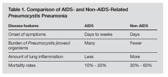
Persons with HIV/AIDS require prophylaxis against PCP when their CD4+ cell count drops below 200/µL or if oral candidiasis develops.5 Patients who are immunosuppressed for other reasons are also vulnerable to PCP. This includes patients with cancer who are undergoing chemotherapy and patients who have received bone marrow or solid organ transplants. In HIV-negative immunosuppressed patients, PCP tends to run a more fulminant course. AIDS-related PCP carries a mortality rate of 10% to 20% for nonintubated patients with a first episode. PCP is more problematic in patients with cancer than in transplant recipients, but mortality from PCP among both of these HIV-negative groups ranges as high as 30% to 60%.6,7 Persons with AIDS have a higher burden of organisms but less inflammation of the alveoli. The more aggressive inflammatory response in persons who are immunosuppressed without AIDS is the cause of more lung injuries and higher mortality rates (Table 1).8

TRANSMISSION OF PCP
It had been assumed that PCP was a reactivation illness in persons with AIDS-as is the case with several other classic opportunistic infections, such as cytomegalovirus retinitis, cerebral toxoplasmosis, and progressive multifocal leukoencephalopathy. Part of this assumption was based on the fact that Pneumocystis is widespread and that 85% of infants have antibodies to the organism by 20 months of age.9
The reactivation concept has not borne out for PCP however, and it is currently believed that PCP is the result of an airborne infection, transmitted from those who are transiently colonized. Colonization implies that the microbe can be isolated in the host but does not cause disease. Pneumocystis organisms have been found in the lungs of both HIV-positive and HIV-negative persons who have not been ill or manifested active infection. Subsequent studies failed to detect the organism, which supported the concept of temporary colonization. Among HIV-negative Pneumocystis hosts, those with chronic lung disease and smokers have higher colonization rates. The colonization rates in HIV-positive patients vary from 10% to 69%.10
Despite the general acceptance of airborne transmission, respiratory isolation of patients with active PCP is not recommended.11
CLINICAL PRESENTATION OF PCP
HIV-positive patients with CD4+ cell counts less than 200/µL who are not receiving prophylaxis may present with a variety of symptoms after PCP develops. Typically, persons with PCP have a dry cough with dyspnea on exertion. These symptoms may have started weeks before. Fever may be present. Other symptoms can include headaches, malaise, night sweats, general fatigue, weight loss, and chest pain. A small percentage (6% to 7%) of persons with PCP are asymptomatic.12,13
Laboratory test results that support a diagnosis of PCP are hypoxia and a widened alveolar-arterial (A-a) gradient. Serum lactate dehydrogenase (LDH) levels tend to be 50 IU/L or more above normal (normal, 90 to 280). In HIV-positive patients with respiratory symptoms and LDH levels above 450 IU/L, PCP is more likely to be the cause than other pulmonary infections.14
Immunosuppressed persons not infected with HIV tend to present with a more acute illness. One early study reported that persons with PCP who were not HIV-infected presented after 5 days of symptoms, whereas those who were HIV-infected presented after 28 days of symptoms.15 Nevertheless, a person with AIDS and PCP can present acutely, and PCP must be considered in all patients who present with clinical evidence of a Pneumocystis infection in the absence of a known HIV test result.
DIAGNOSIS
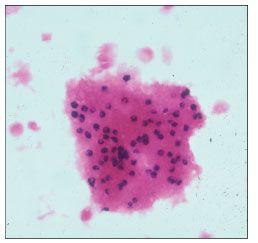
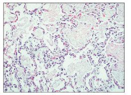
The definitive diagnosis of PCP rests on the identification of P jirovecii organisms obtained in induced sputum or in lung fluid obtained through bronchoalveolar lavage (Figures 1 and 2). Because the diagnostic yield from sputum is highly dependent on the laboratory doing the test, the more reliable procedure remains bronchoscopy. Inability to grow P jirovecii in culture is a persistent obstacle in identifying the infection and assessing potential resistance to treatment.

Figure 1. Bronchial lavage specimen highlighting Pneumocystis jirovecii organisms (Gram Weigart special stain, original magnification ×132). (Photograph courtesy of Dr Amy Chadburn, department of pathology, New York Presbyterian Hospital.)

Figure 2. Histological section of the lung showing "fluffy" eosinophilic infiltrates within the alveolar spaces; these spaces contain Pneumocystis jirovecii organisms. The alveolar walls contain scant inflammatory infiltrates (hematoxylin and eosin stain, original magnification ×33). (Photograph courtesy of Dr Amy Chadburn, department of pathology, New York Presbyterian Hospital.)
Newer techniques that use polymerase chain reaction-based molecular assays show promise but have not yet demonstrated high enough sensitivity and specificity to be used routinely in the clinical setting.15 A 2007 article from Japan reviewed several studies that correlated levels of -D-glucan, a specific component of the Pneumocystis cell wall, with the ability to diagnose PCP.16 In the United States, the fungal cell wall-specifically, the identification of the complex carbohydrates and structural proteins that make up the cell wall-has been a focus of studies of the Pneumocystis disease process and host immune and inflammatory responses. Determination of -D-glucan levels is not yet seen as a means to specifically improve diagnostic reliability.17
Radiology
On chest films, PCP most frequently presents with diffuse, interstitial infiltrates. A 1992 study reviewing 2400 cases reported this ground-glass appearance in nearly 80% of patients with PCP.18
CT is now used routinely for patients with suspected PCP. The interstitial pattern can be central, peripheral, homogeneous, or distributed in a mosaic pattern. Other, less common findings on a CT scan include cavitary lesions, linear opacities, solitary or multiple nodules, cystic formations, and pneumothoraces.16 Gallium scans are used less frequently than CT scans, but they can support a diagnosis of PCP when findings are positive.12
TREATMENT
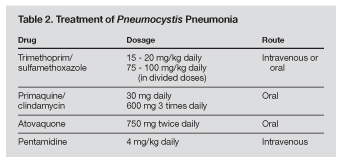
The standard treatment for PCP in HIV-infected persons remains a 21-day regimen of oral or intravenous TMP/ SMX. When indicated, TMP/SMX is given intravenously for at least 7 days before transition to oral therapy. Alternative treatments include intravenous pentamidine, primaquine plus clindamycin, or atovaquone (Table 2). The effectiveness of pentamidine and TMP/SMX in treating PCP is similar, but the potential for adverse effects, such as hypoglycemia and cardiac arrhythmias (torsades de pointes) has limited the use of pentamidine.

Patients with hypoxemia (oxygen pressure below 70 mm Hg) or an A-a gradient of more than 35 mm Hg should also receive corticosteroids. The use of potentially immunosuppressive medication in patients who are already immunodeficient may seem paradoxical to clinicians unfamiliar with treating opportunistic infections in persons with AIDS. However, it is the inflammatory response to the organisms that causes lung injury-not the organisms themselves. The use of corticosteroids is therefore beneficial to patients with acute, severe PCP.
Resistance to TMP/SMX has never been conclusively demonstrated because of the inability to grow the organism in culture. The presence of a mutation in the dihydropteroate synthase (DHPS) gene in other organisms, such as Plasmodium falciparum, Escherichia coli, and Streptococcus pneumoniae, is known to confer resistance to sulfa drugs. This gene and certain nonsynonymous mutations have been sequenced on P jirovecii, but there has not been consistent evidence of treatment failure or success in persons with or without the DHPS mutation.10 Mortality from PCP in HIV-infected persons depends on a variety of factors, but treatment failure as a result of drug resis-tance has not been demonstrated.
PREVENTION/PROPHYLAXIS
All persons who are immunosuppressed should receive prophylaxis against Pneumocystis infection. Those infected with HIV and who have CD4+ cell counts below 200/µL are at the highest risk for PCP when compared with patients immunosuppressed after bone marrow or organ transplant or those receiving cancer chemotherapy.
For patients receiving effective antiretroviral therapy whose CD4+ cell counts recover to levels above 200/µL, PCP prophylaxis can be safely discontinued. HIV RNA levels should be undetectable for more than 3 months before prophylaxis is stopped.19 Those whose CD4+ cell counts fall below 200/µL are once again at risk for P jirovecii infection. Patients who discontinue their medications or whose antiretroviral regimens fail (for any reason) or those who do not receive consistent medical care are vulnerable to a decrease in their T-cell counts. A recent analysis of HIV-positive persons not receiving primary PCP prophylaxis in the United States revealed that these persons fall into 1 of 3 groups20:
• Those who were eligible for prophylaxis but never started.
• Those who were eligible but had stopped before their CD4+ cell counts rose high enough.
• Those who had stopped prophylaxis when their CD4+ cell counts rose above 200/µL but who did not restart when their cell counts fell back below 200/µL.20
In the first category, between 1994 and 2003, the percentage of patients who had a CD4+ cell count below 200/µL, ie, eligible for prophylaxis, but who had never started prophylaxis fell from nearly 50% to approximately 33%. In the second category, the rate of patients who were receiving prophylaxis but stopped it despite the persistence of CD4+ cell counts below 200/µL fell from more than 40% of those eligible in 1994 to approximately 29% in 2003. The third category represented the largest group of patients not receiving indicated PCP prophylaxis in 2003: 38% of patients who had had a rise in their CD4 count owing to effective antiretroviral therapy had not restarted prophylaxis when their CD4+ cell counts fell back under 200/µL.20
The patient in the case presented here is clearly in this last category. He had "fallen off the radar screen" for a long period and had discontinued his medications. Once he discontinued antiretroviral therapy, his CD4+ cell count drifted down to pretreatment levels. This put him at risk once again for contracting PCP. He presented with PCP and was successfully treated with TMP/SMX. He happily agreed to restart antiretroviral therapy, and within several weeks his HIV RNA levels were undetectable. His CD4+ cell count rose to over 100/µL. When his CD4+ cell count stays above 200/µL for 3 months, he will be eligible to stop his PCP prophylaxis.
There are increasingly newer, easier, and more effective regimens available to treat HIV infection. Patients are living longer and longer. However, some patients stop treatment or take their medication incorrectly, and without effective therapy, suppression of virological replication is lost. Bear in mind that patients who were thoroughly adherent to treatment at one point may be less so at another. We as clinicians need to stay vigilant to the immune compromise that can occur with decreases in our patients' CD4+ cell counts. When appropriate, prophylaxis for opportunistic infections must be restarted.
References:
References1. Ball SC. Pneumocystis carinii pneumonia in an untreated HIV-infected patient. AIDS Reader. 2002;12:472-476, 485.
2. Hawksworth DL. When to start HAART for the treatment of HIV infection. Lancet. 2007;7:5-6.
3. Hay JW, Osmond DH, Jacobson MA. Projecting the medical costs of AIDS and ARC in the United States. J Acquir Immune Defic Syndr. 1988;1:466-485.
4. Hoover DR, Saah AJ, Bacellar H, et al. Clinical manifestations of AIDS in the era of Pneumocystis prophylaxis. Multicenter AIDS Cohort Study. N Engl J Med. 1993; 329:1922-1926.
5. Kaplan JE, Masur H, Holmes KK, et al. USPHS/IDSA guidelines for the prevention of opportunistic infections in persons infected with human immunodeficiency virus: an overview. USPHS/IDSA Prevention of Opportunistic Infections Working Group. Clin Infect Dis. 1995;21(suppl 1):S12-S31.
6. Sepkowitz KA, Brown AE, Telzak EE, et al. Pneumocystis carinii pneumonia among patients without AIDS at a cancer hospital. JAMA. 1992;267:832-837.
7. Pareja JG, Garland R, Koziel H. Use of adjunctive corticosteroids in severe adult non-HIV Pneumocystis carinii pneumonia. Chest. 1998;113:1215-1224.
8. Limper AH, Offord KP, Smith TF, et al. Pneumosystis carinii pneumonia. Differences in lung parasite number and inflammation in patients with and without AIDS. Am Rev Respir Dis. 1989;140:1204-1209.
9. Vargas SL, Hughes WT, Santolaya ME, et al. Search for primary infection by Pneumocystis carinii in a cohort of normal, healthy infants. Clin Infect Dis. 2001;32:855-861.
10. Huang L, Morris A, Limper AH, et al. An official ATS workshop summary: recent advances and future directions in Pneumocystis pneumonia (PCP). Proc Am Thorac Soc. 2006;3:655-664.
11. Thomas CF, Limper AH. Pneumocystis pneumonia. N Engl J Med. 2004;350:2487-2498.
12. Barron TF, Birnbaum NS, Shane LB, et al. Pneumocystis carinii pneumonia studied by gallium-67 scanning. Radiology. 1985;154:791-793.
13. Engelberg LA, Lerner CW, Tapper ML. Clinical features of Pneumocystis pneumonia in the acquired immunodeficiency syndrome. Am Rev Respir Dis. 1984;130:689-694.
14. Zaman MK, White DA. Serum lactate dehydrogenase and Pneumocystis carinii pneumonia. Diagnostic and prognostic significance. Am Rev Respir Dis. 1988;137:796-800.
15. Kovacs JA, Hiemenz JW, Macher AM, et al. Pneumocystis carinii pneumonia: a comparison between patients with the acquired immunodeficiency syndrome and patients with other immunodeficiencies. Ann Intern Med. 1984;100:663-671.
16. Fujii T, Nakamura T, Iwamoto A. Pneumocystis pneumonia in patients with HIV infection: clinical manifestations, laboratory findings, and radiological features. J Infect Chemother. 2007;13:1-7.
17. Thomas CF, Limper AH. Current insights into the biology and pathogenesis of Pneumocystis pneumonia. Nat Rev Microbiol. 2007;5:298-308.
18. Kennedy CA, Goetz MB. Atypical roentgenographic manifestations of Pneumocystis carinii pneumonia. Arch Intern Med. 1992;152:1390-1398.
19. Kaplan JE, Masur H, Holmes KK. Guidelines for preventing opportunistic infections among HIV-infected persons-2002. Recommendations of the U.S. Public Health Service and the Infectious Diseases Society of America. MMWR. 2002;51:1-52.
20. Teshale EH, Hanson DL, Wolfe MI, et al. Reasons for lack of appropriate receipt of primary Pneumocystis jiroveci pneumonia prophylaxis among HIV-infected persons receiving treatment in the United States: 1994-2003. Clin Infect Dis. 2007;44:879-883.
2 Commerce Drive
Cranbury, NJ 08512
All rights reserved.