- Clinical Technology
- Adult Immunization
- Hepatology
- Pediatric Immunization
- Screening
- Psychiatry
- Allergy
- Women's Health
- Cardiology
- Pediatrics
- Dermatology
- Endocrinology
- Pain Management
- Gastroenterology
- Infectious Disease
- Obesity Medicine
- Rheumatology
- Nephrology
- Neurology
- Pulmonology
HIV-Associated Pseudotumor Cerebri: A Case Report and Literature Review
Idiopathic intracranial hypertension is a cause of vision loss in HIV-positive patients. In many patients with controlled HIV disease, idiopathic intracranial hypertension develops without any other apparent cause.
Pseudotumor cerebri, idiopathic intracranial hypertension, or benign intracranial hypertension is the syndrome of increased intracranial hypertension in patients without structural brain or cerebrospinal fluid (CSF) abnormalities.1-4 The annual incidence is 0.9 per 100,000 persons.1 Blindness is the most debilitating complication of idiopathic intracranial hypertension, and it occurs more often in patients who do not respond to medical treatment.1 We report a case of idiopathic intracranial hypertension in an HIV-infected person with vision loss who did not respond to therapy.
CASE SUMMARY
A 28-year-old, nonobese, African American man with confirmed HIV infection since 2003 presented to the emergency department (ED) with a 2-week history of headache, vomiting, nausea, and dizziness. He acquired his HIV infection through sexual contact with an HIV-positive man. His disease was well controlled at presentation; his CD4+ cell count was 524/µL and HIV RNA level was below 75 copies/mL. He was being treated with an antiretroviral drug regimen of ritonavir-boosted ata-zanavir, tenofovir, and lamivudine. Findings from a CT scan of the head were unremarkable.
A lumbar puncture was performed in the ED. There was no indication in the patient’s chart that a funduscopic examination had been performed or that a CSF opening pressure had been obtained. CSF studies revealed normal glucose and protein values but a white blood cell (WBC) count of 14/µL (normal, 0 to 6), with 97% lymphocytes (lymphocytic predominance) (normal, 70%). Results of CSF Cryptococcus antigen testing and bacterial, viral, mycobacterial, and fungal cultures were negative at the main hospital laboratory and state reference laboratory. He was discharged on a regimen of promethazine and analgesics for treatment of his symptoms.
The patient returned to the hospital 1 week later with persistent headache, nausea, and vomiting as well as neck stiffness and photophobia. A second lumbar puncture was performed, and the CSF opening pressure was elevated at 430 mm H2O (normal, 60 to 200). New CSF studies revealed a WBC count of 34/µL, with a lymphocytic predominance of 93%, and normal glucose and protein values. Results of repeated CSF Cryptococcus antigen testing and bacterial, viral, mycobacterial, and fungal cultures remained negative. The patient received scheduled analgesics and was discharged.
The patient returned to the HIV clinic with concerns of declining vision, nausea, and headache. A funduscopic examination was performed and showed papilledema. He was admitted to the hospital with a CSF opening pressure of 340 mm H2O on lumbar puncture. Laboratory tests were repeated, and results were unremarkable. Findings from MRI and angiography of the head and spine were unremarkable, and cultures of the CSF and blood were negative. Empiric therapy with acetazolamide was started. His vision improved, and he was discharged.
The patient underwent biweekly lumbar puncture until he was lost to follow-up. He had been nonadherent to his acetazolamide therapy because of diarrhea.
The patient returned to the HIV clinic several months later with decreased vision. An internal CSF shunt was placed, and he underwent optic nerve sheath fenestration but was unable to regain his vision.
DISCUSSION
Idiopathic intracranial hypertension is a diagnosis of exclusion. Criteria for a diagnosis are the following:
• Symptoms and signs attributable to increased intracranial pressure or papilledema.
• Elevated intracranial pressure (greater than 250 mm H2O).
• Normal CSF composition.
• No imaging evidence of ventriculomegaly or a structural cause for increased intracranial pressure.
• No other identified cause of intracranial hypertension.
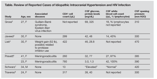
Common symptoms of idiopathic intracranial hypertension are headache; tinnitus; and visual disturbances, including diplopia, visual scotomata, and obscurations. Papilledema and cranial nerve palsies are often observed. Findings from a CSF analysis are usually unremarkable, but occasionally there is a small increase in WBC count and protein level. A CSF culture is usually sterile. Visual loss is typically insidious, but in patients with severe papilledema, the visual loss can progress to permanent blindness within hours. The case of idiopathic intracranial hypertension in our patient is the eighth reported case of the disease in a patient with HIV infection (Table).3-8

The pathogenesis of idiopathic intracranial hypertension remains unclear, but theories revolve around 3 basic principles: increased CSF volume due to excess CSF production, increased cerebral blood volume or brain water content, and obstruction of CSF or venous outflow. CSF lymphocytic pleocytosis has commonly been reported in HIV-infected persons with or without idiopathic intracranial hypertension.1,2 In the literature, non-HIV–related causes, such as cerebrovascular accident, endocrine abnormality, and obesity, are commonly noted to have an association with idiopathic intracranial hypertension.
Therapy is indicated for patients with visual acuity or visual field loss, moderate to severe papilledema, or persistent headaches. Treatment of idiopathic intracranial hypertension may involve CSF removal, weight loss, and surgery. Acetazolamide, the first-line therapy, is a carbonic anhydrase inhibitor that decreases the secretion of CSF by the choroid plexus. Medical treatment is usually given for 6 months in patients who show clinical improvement. One of the following surgical procedures may be used for treatment: optic fenestration, cutting the dura around the optic nerve to decrease pressure, or intracranial shunting to create an artificial passage where excess CSF can be returned to the systemic circulation.
No potential conflict of interest relevant to this article was reported by the authors.
References:
References1. Elovaara I, Iivanainen M, Valle SL, et al. CSF protein and cellular profiles in various stages of HIV infection related to neurological manifestations. J Neurol Sci. 1987;78:331-342.
2. Hollander H, Stringari S. Human immunodeficiency virus-associated meningitis. Clinical course and correlations. Am J Med. 1987;83:813-816.
3. Gross FJ, Mindel JS. Pseudotumor cerebri and Guillain-Barré syndrome associated with human immunodeficiency virus infection. Neurology. 1991;41: 1845-1846.
4. Javeed N, Shaikh J, Jayaram S. Recurrent pseudotumor cerebri in an HIV-positive patient. AIDS. 1995; 9:817-819.
5. Lisk DR, Cummings CC, Charles CC, et al. Rapid weight gain and benign intracranial hypertension in an AIDS patient on treatment with highly active anti-retroviral therapy (HAART). West Indian Med J. 2000;49:338-339.
6. Prevett MC, Plant GT. Intracranial hypertension and HIV associated meningoradiculitis. J Neurol Neurosurg Psychiatry. 1997;62:407-409.
7. Schwarz S, Husstedt IW, Georgiadis D, et al. Benign intracranial hypertension in an HIV-infected patient: headache as the only presenting sign. AIDS. 1995;9:657-658.
8. Traverso F, Stagnaro R, Fazio B. Benign intracranial hypertension associated with HIV infection. Eur Neurol. 1993;33:191-192.
