- Clinical Technology
- Adult Immunization
- Hepatology
- Pediatric Immunization
- Screening
- Psychiatry
- Allergy
- Women's Health
- Cardiology
- Pediatrics
- Dermatology
- Endocrinology
- Pain Management
- Gastroenterology
- Infectious Disease
- Obesity Medicine
- Rheumatology
- Nephrology
- Neurology
- Pulmonology
Young Child With Hematuria and Dysuria
A 3-year-old girl is brought to the office because of a1-week history of hematuria and dysuria. Her motherhad noticed bright red blood in the child’s urine anddiaper. The child did not have dysuria initially but latercomplained of a burning sensation.

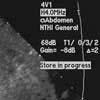
Figure 1

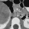
Figure 2
A 3-year-old girl is brought to the office because of a 1-week history of hematuria and dysuria. Her mother had noticed bright red blood in the child's urine and diaper. The child did not have dysuria initially but later complained of a burning sensation. A week earlier, the patient had been seen at an urgent care center. Oral trimethoprim/sulfamethoxazole was prescribed after urinalysis showed numerous red blood cells and few white blood cells. However, the hematuria persisted.
History. The child was delivered vaginally at full term and has had normal developmental milestones. Her immunizations are up-to-date. The child was previously healthy, and she has had no recent contact with ill persons.
The family history is noncontributory. The patient has no allergies and takes no medications regularly.
Examination. This well-built, well-nourished, afebrile child is smiling and appears happy. Weight, 30 lb. Height, 36.5 inches. Heart rate, 110 beats per minute and regular; respiration rate, 22 breaths per minute; blood pressure (BP), right upper limb, 145/110 mm Hg; left upper limb, 142/110 mm Hg; right lower limb, 138/106 mm Hg. She is well hydrated. Examination of the head and neck shows no erythema, icterus, edema, or evidence of candidal infection. There is no palpable lymphadenopathy, joint abnormality, or ankle edema. Abdominal examination reveals no organomegaly or tenderness. Bowel sounds are normal. Hernial orifices are clear; genital examination is normal. The jugular vein pulse is normal and the apex is within normal limits. Both heart sounds are heard with S4. All peripheral pulses are palpable. Lungs are clear. Results of the neurological and funduscopic examination are unremarkable.
Laboratory studies. Hemoglobin, 13.6 g/dL; white blood cell (WBC) count, 8500/µL with 42% lymphocytes, 52% segmented neutrophils, 2% eosinophils, and 4% monocytes. Erythrocyte sedimentation rate, 8 mm/h. Urinalysis reveals specific gravity, 1.050; protein, 100 mg/dL; no glucose; leukocyte esterase, negative; red blood cells, 280/µL; WBC, 29/µL. Gram staining is negative for bacteria; culture is negative for pathogens. Total bilirubin, 0.2 mg/dL; alkaline phosphatase, 182 U/L; aspartate aminotransferase, 40 U/L; alanine aminotransferase, 38 U/L. Serum sodium, 137 mEq/L; potassium, 3.9 mEq/L; chloride, 101 mEq/L; carbon dioxide,22 mEq/L. Serum glucose, 92 mg/dL; blood urea nitrogen, 11 mg/dL; creatinine, 0.4 mg/dL.
An ultrasound scan of the kidney and an abdominal CT are ordered.
Based on the clinical picture and the imaging studies, what is the most likely diagnosis?
A. Renal abscess
B. Wilms tumor (nephroblastoma)
C. Nephrolithiasis with hydronephrosis
D. Acute glomerulonephritis
WILMS TUMOR: AN OVERVIEW
Wilms tumor is the most common primary malignancy of the kidney and the second most common abdominal tumor of childhood. It represents 5% to 7% of childhood cancers in the United States. Ninety percent of children present before the age of 7 years; the peak incidence is between 3 and 4 years. It affects boys and girls equally; the incidence is higher in African American children than in white or Asian children. Familial inheritance accounts for 1% of cases.
PATHOGENESIS AND GENETICS
Wilms tumor is thought to arise from abnormal proliferation of pluripotent embryonic renal precursor cells that fail to undergo proper differentiation into tubules and glomeruli. These cell populations are present in 1 of 100 neonatal postmortem examinations; however, the overall incidence of Wilms tumor is 1 in 10,000, which suggests that only 1% undergo malignant conversion.
Three genetic conditions are associated with Wilms tumor:
- Wilms tumor, Aniridia, Genitourinary malformation, and mental Retardation (WAGR) syndrome.
- Denys-Drash syndrome (Wilms tumor, male pseudohermaphroditism, renal mesangial sclerosis).
- Beckwith-Wiedemann syndrome (large birthweight, hemihypertrophy, macroglossia, hepatomegaly, and predisposition to Wilms tumor and other malignant disorders).
The risk of Wilms tumor in patients with WAGR or Denys-Drash syndrome is about 30%; overgrowth syndromes, such as Beckwith-Wiedemann, carry an estimated risk of 10%.
Two major gene loci are involved in the development of Wilms tumor. WT1, which is located on chromosome 11p13, is linked to both WAGR and Denys-Drash syndromes. WT1 encodes for a transcription factor involved in normal kidney and gonadal development. WT2, located on chromosome 11p15.5, is associated with Beckwith-Wiedemann syndrome. The role of the WT2 gene remains to be defined.
DIAGNOSIS
Wilms tumor is most frequently detected after an asymptomatic abdominal mass is palpated. Other presenting features may include chronic abdominal pain, anorexia, vomiting, malaise, hematuria, and hypertension. Less frequent manifestations, such as an acute abdomen secondary to tumor rupture or a varicocele from tumor invasion into the inferior vena cava, have also been documented.
Because of the increased risk of Wilms tumor in children with aniridia, hemihypertrophy, or Beckwith-Wiedemann syndrome, routine screening with ultrasonography every 3 months is recommended until age 7 years, followed by semiannual physical examinations until somatic growth is complete. Some experts recommend the use of this protocol with children who have early-onset nephrotic syndrome that involves diffuse mesangial sclerosis, with or without ambiguous genitalia.
Confirmation of the diagnosis begins with Doppler ultrasonography to identify the source of the mass, identify any genitourinary abnormalities, confirm the presence of a contralateral kidney, and determine whether the tumor extends into the inferior vena cava. CT with or without contrast can detect sharp demarcations between tumor and healthy renal parenchyma. MRI offers further refinement of the anatomical configuration by producing multiplanar views of the kidney and abdomen with superior soft tissue contrast.
Additional imaging options include chest radiography to identify possible metastatic disease; the lungs are the most common site of metastasis. The need for chest CT in patients with normal chest radiographs is controversial; it is unclear whether lesions detected by CT alone require more aggressive treatment.
Definitive diagnosis and staging are achieved via tissue biopsy, which is done at the time of resection. A biopsy is not typically performed before surgery (except as discussed below) because of the risk of tumor leakage from the puncture site or seeding of the needle tract, which may result in relapse.
The staging of Wilms tumor is as follows:
- Stage I: Tumor is confined to the kidney and is completely resected.
- Stage II: Tumor extends beyond the kidney (eg, from penetration of the renal capsule, invasion of the renal sinus vessels, biopsy before tumor removal, or spillage of the tumor locally during removal) but is ultimately completely resected with negative margins and lymph nodes.
- Stage III: Gross or microscopic tumor remains after surgery.
- Stage IV: Hematogenous or lymph node metastases.
- Stage V: Bilateral Wilms tumors.
Histological assessment is also undertaken at this point. In conjunction with staging, this evaluation guides therapy and provides the most important indicators for prognosis.
MANAGEMENT
Treatment involves surgery, chemotherapy, and/or radiation. Initial management is nearly always surgical and consists of unilateral nephrectomy. Chemotherapy is typically initiated after surgery and is administered for 18 to 24 weeks. Generally, stage I and stage II disease with favorable histology are treated with vincristine and dactinomycin; doxorubicin is added for stage III and stage IV disease.
Therapy for unfavorable histology tumor types, including diffuse anaplasia, clear cell sarcoma of the kidney, and rhabdoid tumor of the kidney, involves etoposide, cyclophosphamide, and/or carboplatin. Radiation is administered for all tumor types except stage I with favorable or anaplastic histology and stage II with favorable histology.
For patients with stage V Wilms tumors, solitary kidney, intravascular extension of the tumor above the hepatic veins, or inoperable disease at presentation, neoadjuvant chemotherapy is initiated after biopsy (without resection) in hopes of shrinking the tumor and facilitating renal-sparing procedures. Surgery (if possible) is usually performed no later than 12 to 18 weeks after initial diagnosis. Additional chemotherapy or radiation postoperatively may also be required.
PROGNOSIS
The 4-year survival for patients with favorable histology Wilms tumor approaches 90% when the tumor is localized and 80% in patients with metastatic disease. The survival rate among patients with stage V Wilms tumors exceeds 70%; however, renal failure develops in many of these persons.
LONG-TERM EFFECTS
Long-term survivors require monitoring. Recurrence occurs most often in the first 2 years after nephrectomy but has been documented as late as 11 years postoperatively. The rate of secondary malignancies is 6% at 20 years; irradiated areas are the primary sites affected.
Another potential late effect is end-stage renal disease, which is most common in patients with Denys-Drash syndrome or those who have undergone bilateral nephrectomy. Specific late effects of radiation include scoliosis, ovarian failure, oligospermia, azoospermia, and decreased cardiac or pulmonary function, depending on which organs were exposed. Chemotherapy-related side effects include heart failure (secondary to doxorubicin), hepatotoxicity (from dactinomycin or combination therapy), nephrotoxicity (from etoposide), hemorrhagic cystitis (from cyclophosphamide if not used with mesna), infertility, and secondary malignancies (leukemias).
Current treatment protocols recommend lower dosages of chemotherapy and radiation for low-risk patients, with the hope of reducing the prevalence of late effects without sacrificing the excellent cure rates.
References:
FOR MORE INFORMATION:
- Jones KP. Controversies and advances in the management of Wilms' tumor. Arch Dis Childhood. 2002;87:241-244.
- Kalapurakal JA, Dome JS, Perlman EJ, et al. Management of Wilms' tumor: current practice and future goals. Lancet Oncol. 2004;5:37-46.
- Keaney CM, Springate JE. Cancer and the kidney. Adolesc Med Clin. 2005;16: 121-148.
- Neville HL, Ritchey ML. Wilms' tumor: overview of National Wilms' Tumor Study Group results. Urol Clin North Am. 2000;27:435-442.
- Ozcan T, Bahado-Singh R. Prenatal diagnosis of fetal renal masses. UpToDate. 2005.
- Weiner JS, Coppes MJ, Ritchey ML. Current concepts in the biology and management of Wilms' tumor. J Urol. 1998;159:1316-1325.
- Wu HY, Snyder HM 3rd, D'Angio GJ. Wilms' tumor management. Curr Opin Urol. 2005;15:273-276.
