- Clinical Technology
- Adult Immunization
- Hepatology
- Pediatric Immunization
- Screening
- Psychiatry
- Allergy
- Women's Health
- Cardiology
- Pediatrics
- Dermatology
- Endocrinology
- Pain Management
- Gastroenterology
- Infectious Disease
- Obesity Medicine
- Rheumatology
- Nephrology
- Neurology
- Pulmonology
Woman With Edema, Vomiting, and Diarrhea
A 22-year-old woman has had chronic nausea, emesis with green vomitus, and diarrhea for the past 10 months. The diarrhea is frequent (about 3 to 8 times daily) and does not resolve with starvation.

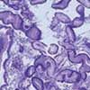
Figure 1

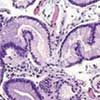
Figure 2
A 22-year-old woman has had chronic nausea, emesis with green vomitus, and diarrhea for the past 10 months. The diarrhea is frequent (about 3 to 8 times daily) and does not resolve with starvation. The stools are loose to watery and foul-smelling; there is no mucus or pus. She has also had progressive pedal edema over the past month.
The patient has had no abdominal or chest pain, hematemesis, hematochezia, melena, jaundice, polyuria, skin infection, upper respiratory tract infection, hematuria, shortness of breath, cough, orthopnea, or fever. She has a 3-pack-year smoking history but has recently quit. The patient's mother had noticed facial puffiness about a year before the onset of her other symptoms.
The mother has rheumatoid arthritis. The patient's sister has systemic lupus erythematosus, which was diagnosed at age 17 years.
The patient has bilateral grade 4 pitting pedal edema that extends up to the thighs and abdominal wall edema. The neck is supple with no elevated jugular venous pressure or lymphadenopathy. Breath sounds are decreased at the bases on both sides. There are multiple striae on the abdomen but no shifting dullness or fluid thrill. The remainder of the physical findings are unremarkable.
Review of laboratory studies performed during the past several months reveals low levels of albumin (0.4 g/dL) and total protein (2.4 g/dL). Results of a urinalysis, 24-hour urinary protein measurement, liver function tests, liver ultrasonography, and transthoracic echocardiography are normal. A pregnancy test is negative. Stool studies are negative for leukocytes, Giardia, and Clostridium difficile toxin. Fecal fat (neutral and split) is normal. The α1-antitrypsin level is elevated to 0.79 mg/g of stool (normal, 0 to 0.62 mg/g of stool).
The serum α1-antitrypsin level is 42 mg/dL (normal,83 to 199 mg/dL). Serum protein levels are also below normal. Serum IgG is 195 mg/dL (normal, 636 to 1300 mg/dL); IgA and IgM are within the normal range. Tests for tissue antitransglutaminase antibody, anti-endomysial antibody, and antigliadin antibody are negative. Antinuclear antibody is positive (titer of 1:320); anti–double-stranded DNA and anti-Smith antibodies are negative. Results of a colonoscopy with biopsy and a small-intestine biopsy are normal.
Esophagogastroduodenoscopy (EGD) reveals giant, enlarged gastric rugae. Biopsy specimens are shown.
What is the most likely cause of this patient's symptoms?
- Celiac disease
- Whipple disease
- Lymphangiectasia
- Zollinger-Ellison syndrome
- Mntrier disease
- Inflammatory bowel disease
- Nephrotic syndrome
(Answer on the next page.)

Figure 1

Figure 2
Mntrier disease
Histopathological examination revealed mild to moderate foveolar hyperplasia and decreased parietal cells. These biopsy results and the finding of giant, hypertrophic gastric folds on the EGD confirmed the diagnosis of Mntrier disease.
DIFFERENTIAL DIAGNOSIS
Celiac disease was ruled out on the basis of the laboratory workup, which was negative for antitransglutaminase, antigliadin, and anti-endomysial antibodies, and the intestinal biopsy findings, which showed no generalized villous atrophy.
Whipple disease was ruled out because the intestinal biopsy findings showed no periodic acid-Schiff–positive macrophages.
Lymphangiectasia would appear as dilated lymphatic vessels, especially at the tip of the villi, on the intestinal biopsy.
Zollinger-Ellison syndrome (characterized by an increased number of parietal cells) also causes hyperplastic gastropathy; however, the finding of decreased parietal cells on the gastric biopsy ruled out this condition.
Inflammatory bowel disease was excluded by the normal colonoscopy/biopsy results.
Nephrotic syndrome was ruled out by the normal 24-hour urinary protein level.
The differential diagnosis in any patient with anasarca includes cardiac (heart failure), renal (nephrotic syndrome), nutritional (hypoproteinemia), and hepatic (cirrhosis) causes. This patient's history of nausea, vomiting, and diarrhea suggested protein-losing enteropathy.
Protein-losing enteropathy encompasses a group of conditions that are characterized by excessive loss of proteins in the GI tract, which causes hypoproteinemia and edema. The pathology results from excessive protein excretion or defects in absorption.
The initial test for a suspected protein-losing enteropathy is the measurement of stool a1-antitrypsin, which is elevated with a decrease in serum proteins across the spectrum, rather than selective loss. In a subgroup of patients, the cause is a malabsorption syndrome, such as celiac disease, tropical sprue, chronic pancreatitis, or inflammatory bowel disease. Because the causes of protein-losing enteropathy are multiple and varied, an algorithmic approach is required to establish the diagnosis. The workup includes, but is not limited to, panendoscopy.
MNTRIER DISEASE: AN OVERVIEW
Mntrier disease-also known as hypertrophic gastritis, protein-losing gastropathy, hyperplastic or hypertrophic gastropathy, and polyadenomes polypeux-is very rare. Cytomegalovirus (CMV) infection, Helicobacter pylori infection, abnormalities in the epidermal growth factor receptor, and transforming growth factor a overexpression are suggested causes. Symptoms include epigastric pain accompanied by nausea, vomiting, anorexia, and weight loss. Occult GI bleeding may occur. However, overt bleeding is unusual and when present is a result of superficial mucosal erosions. Anasarca may be the presenting sign.
An upper GI series may show enormous filling defects that can be mistaken for malignancy. EGD shows large, tortuous gastric mucosal folds-the classic feature of the disease-most prominent in the body and fundus of the stomach. Consider Mntrier disease only in patients with documented protein-losing enteropathy and large gastric folds on EGD. The differential diagnosis for large gastric folds includes Zollinger-Ellison syndrome, malignancy, infection (CMV infection, histoplasmosis, syphilis), and infiltrative disorders (such as sarcoidosis). Deep mucosal biopsy (and cytology) is required to establish the diagnosis of Mntrier disease.
Treatment includes a high-protein diet, anticholinergic agents, prostaglandins, proton pump inhibitors, prednisone, H2-receptor antagonists, anti–H pylori therapy, and octreotide. No treatment has proved to be consistently effective. Severe disease with persistent and substantial protein loss may require total or subtotal gastrectomy.
OUTCOME OF THIS CASE
This patient was given symptomatic treatment with furosemide and antiemetics and an albumin infusion. Therapy for suspected H pylori infection was also provided. A high-protein diet containing medium-chain triglycerides was recommended.
The patient required total parenteral nutrition during most of her hospital stay. She died after 5 months of complications associated with catheter-related sepsis.
