- Clinical Technology
- Adult Immunization
- Hepatology
- Pediatric Immunization
- Screening
- Psychiatry
- Allergy
- Women's Health
- Cardiology
- Pediatrics
- Dermatology
- Endocrinology
- Pain Management
- Gastroenterology
- Infectious Disease
- Obesity Medicine
- Rheumatology
- Nephrology
- Neurology
- Pulmonology
Using ultrasonography in the diagnosis and management of pleural disease
ABSTRACT: The increasing availability of bedside ultrasonographyallows for more timely diagnosis and treatment of pleuraleffusion while limiting the patient's exposure to radiation. Thedynamic signs characteristic of pleural effusions includerespirophasic changes in the shape of the fluid collection, floatingmovements of atelectatic lung, and the plankton sign. Ultrasonographyalso is an efficient means of excluding pneumothoraxwhen rapid diagnosis is needed or after interventionssuch as central line placement, lung or pleural biopsy, or thoracentesis.The diagnosis of a pneumothorax relies on the absenceof dynamic signs such as "lung sliding." Static signs, suchas the comet tail artifact, or consolidated lung parenchyma orlung tissue that contains a solid mass, also can be useful in excludingpneumothorax. Ultrasonography can be used to guidefine-needle aspiration and core biopsies of pleural nodules,pleural thickening, and subpleural lung masses. (J Respir Dis.2008;29(5):200-207)
ABSTRACT: The increasing availability of bedside ultrasonography allows for more timely diagnosis and treatment of pleural effusion while limiting the patient's exposure to radiation. The dynamic signs characteristic of pleural effusions include respirophasic changes in the shape of the fluid collection, floating movements of atelectatic lung, and the plankton sign. Ultrasonography also is an efficient means of excluding pneumothorax when rapid diagnosis is needed or after interventions such as central line placement, lung or pleural biopsy, or thoracentesis. The diagnosis of a pneumothorax relies on the absence of dynamic signs such as "lung sliding." Static signs, such as the comet tail artifact, or consolidated lung parenchyma or lung tissue that contains a solid mass, also can be useful in excluding pneumothorax. Ultrasonography can be used to guide fine-needle aspiration and core biopsies of pleural nodules, pleural thickening, and subpleural lung masses. (J Respir Dis. 2008;29(5):200-207)
Ultrasonography is an important modality in the evaluation of pleural disease. The increasing availability of portable ultrasonography units in clinical areas, such as procedure suites, emergency departments, and ICUs, has made ultrasound equipment accessible to many physicians involved in the treatment of patients with pleural pathology.
However, as sophisticated as modern hand-carried ultrasonography units are and as good as image quality has become, an understanding of the principles of ultrasonography and image interpretation has to be brought to the bedside by the physician. Fortunately, pleural ultrasonography poses few challenges to the beginning physician-sonographer. This makes it ideally suited, with vascular access sonography, for the enterprising novice, especially when compared with limited echocardiography or abdominal ultrasonography.
The primary challenges of pleural sonography are the identification of pleural effusion as well as the safe and successful performance of drainage procedures. Pleural ultrasonographic findings and uses are summarized in Table 1. A glossary of the relevant terms is presented in Table 2.
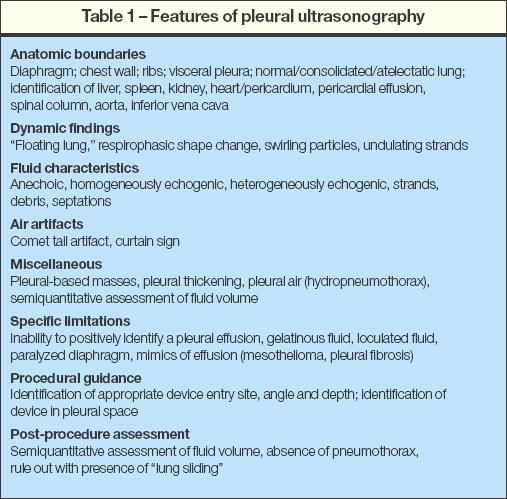
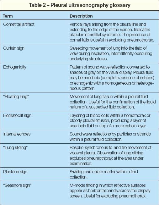
In this article, we will review the use of ultrasonography in the evaluation of pleural effusions and pneumothorax and the role of interventional ultrasonography in patients with pleural disease.
THE BASICS
Ultrasound hardware
In recent years, high-resolution portable ultrasonography units have become ubiquitous in the hospital setting. In general, sonography machines that are capable of abdominal ultrasonography or echocardiography are also suitable for pleural sonography. Specifically, convex array or sector scanning ultrasound probes with frequencies between 3 and 5 MHz provide a good balance between near-field resolution and depth penetration. Transducers that have higher frequencies are more suitable for the evaluation of the chest wall. Color Doppler is of limited use in pleural ultrasonography.
There is no standardized operator interface for sonography machines except for an adjustable marker on the screen that corresponds to a ridge or groove on the transducer. By convention, the marker is placed on the left upper corner of the ultrasound image, and the marker on the probe points cephalad.
Technical aspects of image acquisition
Patients able to sit upright should be placed in the same position as for thoracentesis but with enough space between the anterior chest and the arm support to enable anterior scanning. Patients incapable of sitting upright, especially those who are critically ill, may be examined in whichever position can be achieved with 1 or more assistants holding the patient, if required.
The ultrasound probe is applied to the chest wall so that the marker is pointed cephalad, and rib inter-spaces are examined in a longitudinal fashion. The probe is then moved medially or laterally, and another longitudinal examination is performed. This allows for an examination of the entire costal pleura. The mediastinal pleura is not readily evaluated by transthoracic ultrasonography.1
DIAGNOSTIC ULTRASONOGRAPHY
Pleural effusion
A pleural effusion is usually suspected on the basis of findings from the physical examination or a standard chest radiograph. Sonography can be used to confirm the presence or absence of pleural effusion (Table 3).

The diagnosis of pleural effusion can be made when an echo-free fluid collection within the boundaries of the parietal and visceral pleurae is visualized and any of the dynamic signs characteristic of pleural effusions are observed. These signs include respirophasic changes in the shape of the fluid collection, floating movements of atelectatic lung ("floating lung"), undulating movements of fibrinous strands, and a swirling motion of debris (plankton sign).
The boundaries of a pleural effusion are defined by the identification of organs adjacent to the effusion- in particular, the liver, spleen, heart, and lung (Figure 1). The lung adjacent to a pleural effusion may be atelectatic, consolidated, or aerated. An aerated lung can be visualized only indirectly by the typical air artifacts, such as comet tails, or total reflection of the ultrasound beam with obscuration of underlying structures.
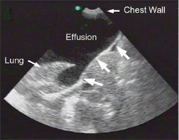
Figure 1 –
This anechoic pleural effusion is contained within the defining anatomic boundaries, such as the lung, chest wall, and diaphragm (large arrows).
The border between atelectatic and aerated lung is often visible, and progressive expansion of aerated lung can be observed during thoracentesis. Ultrasonography may reveal lung tumors or pleural nodules and masses and can easily distinguish these from a coexisting pleural effusion (Figure 2).2-4
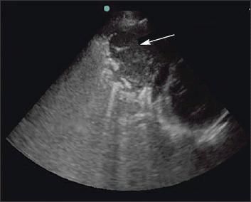
Figure 2 –
A pleuralbased left lung mass can be seen on this ultrasonogram (arrow). Ultrasonography can easily distinguish tumors from coexisting pleural effusion.
Although the presence of an anechoic structure suggests fluid, the absence of echogenicity can also occur with lymphomas, mesothelioma, pleural fibrosis, and highly viscous matter. An anechoic pleural effusion may be either a transudate or an exudate, while the presence of internal echoes in a pleural effusion indicates with a high probability that the fluid is an exudate.5
Complex pleural effusions have internal echoes that may be diffusely distributed within the fluid or may be in a septate pattern (Figure 3). A complex septate pattern is caused by fibrin strands in the fluid and may indicate a complicated parapneumonic effusion, empyema,m or hemothorax, but it does not necessarily indicate loculation.
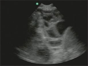
Figure 3 –
Complex pleural effusions may occur in a septate pattern, as seen here, or they may be diffusely distributed within the fluid. A complex septate pattern is caused by fibrin strands in the fluid and may indicate the presence of a complicated parapneumonic effusion, empyema, or hemothorax, but it does not necessarily indicate loculation.
We usually elect to insert a small-bore catheter for drainage of sonographically complex effusions because, in our experience, successful drainage is unlikely with standard thoracentesis catheters. The eventual need for surgical intervention cannot be predicted by the type of internal echoes visualized by ultrasonography.6,7 Hemothoraces may also demonstrate internal echoes with an anechoic phase overlying a homogeneously echogenic phase (hematocrit sign).8
Pneumothorax
Ultrasonography is an efficient means of excluding pneumothorax when rapid diagnosis is needed or after interventions such as central line placement, lung or pleural biopsy, or thoracentesis (Table 4). The patient also may be spared additional radiation exposure.9
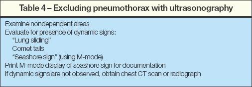
Because ultrasound waves are totally reflected by air, pneumothoraces cannot be directly observed. The diagnosis of a pneumothorax relies on the absence of dynamic signs such as lung movement with respirations, which is seen as a to-and- fro movement of the visceral pleura; this is called "lung sliding." The presence of this sign virtually excludes a pneumothorax at the point of observation.10
Because air accumulates in a nondependent manner, examination of the anterior chest of the supine patient is optimal for the exclusion of pneumothorax. Using this technique, the absence of lung sliding has a sensitivity that is comparable to the sensitivity of CT scanning. Documentation of lung sliding for the medical record can be accomplished by switching the ultrasound to motion mode (M-mode) and observing the "seashore sign" (Figure 4).
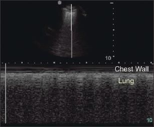
Figure 4 –
This split-screen display of pleural ultrasonography includes the normal display (top) and M-mode display with "seashore sign" (bottom).
Static signs, such as the comet tail artifact, or the observation of consolidated lung parenchyma or lung tissue that contains a solid mass also can be useful in excluding pneumothorax (Figure 5).11 The presence of lung sliding or comet tails is highly sensitive for the absence of anterior pneumothorax in the supine patient. However, if these signs are not present, confirmation by either a chest radiograph or a CT scan is recommended.
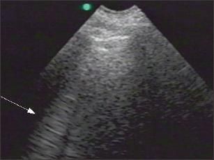
Figure 5 –
The comet tail artifact (arrow) is an example of a static sign that can be useful in excluding pneumothorax.
INTERVENTIONAL ULTRASONOGRAPHY
Thoracentesis
Traditionally, lateral decubitus chest radiographs have been used to confirm the presence of pleural fluid and to document the free-flowing nature of the effusion. Such free-flowing effusions are considered amenable to thoracentesis by the clinician. Patients with loculated pleural effusions are often referred to interventional radiology for drainage.
The increasing availability of bedside ultrasonography allows the pulmonologist or internist to localize and evaluate a pleural fluid collection immediately; in addition, it allows for procedural guidance so that thoracentesis can be performed in a timely fashion by the clinician without the need for referral to interventional radiology. As a result, the use of bedside ultrasonography generally allows for more timely diagnosis and treatment of pleural effusion while limiting the patient's exposure to radiation.12
Ultrasonography provides important information about the quantity, location, and character of pleural fluid. The use of image guidance for thoracentesis may increase the likelihood of successful thoracentesis and may decrease the incidence of complications.13-16 A study comparing ultrasonographically selected thoracentesis sites with sites that were chosen on the basis of physical examination findings demonstrated that clinically chosen sites would have resulted in the puncture of solid organs, such as the liver or spleen, in up to 15% of cases.17
Sonographic localization of the pleural fluid should be performed immediately before thoracentesis and without any changes in the patient's position, since free-flowing fluid will change locations as the patient moves.1 Once the location of the effusion is identified with ultrasound, a mark corresponding to the largest area of fluid collection is made on the patient's skin, and the thoracentesis is performed using standard technique.
Because of the ease of performance, higher success rate, and improved safety, imageguided thoracentesis should be performed if this technology is readily available. Ultrasoundguided thoracentesis has been shown to be safe in patients who are mechanically ventilated, and thoracentesis should always be performed by this method in these patients.1,11 Thoracentesis of small effusions, loculated fluid, or fluid that is not free-flowing should be attempted only under image guidance.
Image-guided catheter placement
The use of image-guided, small-bore drainage catheters for the management of empyemas or malignant effusions is associated with fewer complications (such as hemorrhage), fewer tube malpositions, and less patient discomfort than are largebore thoracostomy tubes. Sonography allows for real-time imaging of the catheter tip or guidewire and is preferred to CT for unstable or uncooperative patients (Figure 6).7,13 Because air reflects ultrasound waves and prevents visualization of intrapleural air, ultrasonography is not a reliable way to identify safe catheter insertion sites in a pneumothorax.
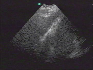
Figure 6 – Sonography allows for real-time imaging of the catheter tip or guidewire and is preferred to CT for unstable or uncooperative patients. Here, a J-wire is visualized after placement into the pleural space.
Ultrasonographically guided biopsy
Ultrasonography can be used to guide fine-needle aspiration and core biopsies of pleural nodules, pleural thickening, and subpleural lung masses. Ultrasound guidance is less invasive than thoracoscopy and does not require general anesthesia.
Direct visualization of pleural thickening or nodules improves diagnostic yield over that of blind pleural biopsy.18,19 Pleural effusions need not be present for fine-needle aspiration or core biopsy of a pleural nodule to be performed using this technique.
HOW TO GET STARTED
The obvious starting point for the beginning thoracic sonographer is the incorporation of sonographic imaging into the thoracentesis procedure. In our experience, a thorough review of narrated cases on video is sufficient for the physician who is already performing thoracentesis regularly. Review of a DVD dedicated to ultrasonography for pleural effusions is now standard in our pulmonary fellowship program at the Medical University of South Carolina.20 Only after physicians review this resource do we allow independent use of sonographic guidance of thoracentesis. We also recommend that physicians review an atlas of thoracic sonography.21
CONCLUSION
Generally, the internist, pulmonologist, surgeon, or emergency physician can easily learn ultrasonography performance and interpretation skills. Recent advances in technology have resulted in wider availability of affordable, portable ultrasonography machines. Increasingly, ultrasonography is considered to be an extension of the physical examination and is used to provide guidance for venous access, thoracentesis, biopsies, and a number of other procedures.
Examination of the pleural space for effusions and pneumothorax is a straightforward application that can be easily learned by clinicians. Ultrasonography of the pleural space provides guidance for invasive procedures, enabling physicians to perform the procedures safely and independently. It is expected that the use of ultrasonography by clinicians will continue to expand as its importance becomes more widely recognized.
References:
REFERENCES
1. Mayo PH, Doelken P. Pleural ultrasonography.
Clin Chest Med.
2006;27:215-227.
2. Lin MS, Hwang JJ, Chong IW, et al. Ultrasonography of chest diseases: analysis of 154 cases.
Gaoxiong Yi Xue Ke Xue Za Zhi.
1992;8:525-534.
3. Lomas DJ, Padley SG, Flower CD. The sonographic appearances of pleural fluid.
Br J Radiol.
1993;66:619-624.
4. Marks WM, Filly RA, Callen PW. Real-time evaluation of pleural lesions: new observations regarding the probability of obtaining free fluid.
Radiology.
1982;142:163-164.
5. Hirsch JH, Rogers JV, Mack LA. Real-time sonography of pleural opacities.
AJR.
1981;136:297-301.
6. Kearney SE, Davies CW, Davies RJ, Gleeson FV. Computed tomography and ultrasound in parapneumonic effusions and empyema.
Clin Radiol.
2000;55:542-547.
7. Shankar S, Gulati M, Kang M, et al. Imageguided percutaneous drainage of thoracic empyema: can sonography predict the outcome?
Eur Radiol.
2000;10:495-499.
8. Yang PC, Luh KT, Chang DB, et al. Value of sonography in determining the nature of pleural effusion: analysis of 320 cases.
AJR.
1992;159:29-33.
9. Maury E, Guglielminotti J, Alzieu M, et al. Ultrasonic examination: an alternative to chest radiography after central venous catheter insertion?
Am J Respir Crit Care Med.
2001;164:403-405.
10. Lichtenstein DA, Menu Y. A bedside ultrasound sign ruling out pneumothorax in the critically ill. Lung sliding.
Chest.
1995;108:1345-1348.
11. Lichtenstein D, Mezière G, Biderman P, Gepner A. The comet-tail artifact: an ultrasound sign ruling out pneumothorax.
Intensive Care Med.
1999;25:383-388.
12. Lipscomb DJ, Flower CD, Hadfield JW. Ultrasound of the pleura: an assessment of its clinical value.
Clin Radiol.
1981;32:289-290.
13. O'Moore PV, Mueller PR, Simeone JF, et al. Sonographic guidance in diagnostic and therapeutic interventions in the pleural space.
AJR.
1987;149:1-5.
14. Weingardt JP, Guico RR, Nemcek AA Jr, et al. Ultrasound findings following failed, clinically directed thoracenteses.
J Clin Ultrasound.
1994;22:419-426.
15. Grogan DR, Irwin RS, Channick R, et al. Complications associated with thoracentesis. A prospective, randomized study comparing three different methods.
Arch Intern Med.
1990;150:873-877.
16. Lichtenstein D, Hulot JS, Rabiller A, et al. Feasibility and safety of ultrasound-aided thoracentesis in mechanically ventilated patients.
Intensive Care Med.
1999;25:955-958.
17. Diacon AH, Brutsche MH, Solèr M. Accuracy of pleural puncture sites: a prospective comparison of clinical examination with ultrasound.
Chest.
2003;123:436-441.
18. Maskell NA, Gleeson FV, Davies RJ. Standard pleural biopsy versus CT-guided cutting-needle biopsy for diagnosis of malignant disease in pleural effusions: a randomized controlled trial.
Lancet.
2003;361:1326-1330.
19. Heilo A, Stenwig AE, Solheim OP. Malignant pleural mesothelioma: US-guided histologic coreneedle biopsy.
Radiology.
1999;211:657-659.
20. Doelken P, Mayo P.
Ultrasonography of Pleural Effusion
[DVD]. Charleston, SC: DM&C Electronic Publishing; 2003.
21. Mathis G, Lessnau KD, eds.
Atlas of Chest Sonography.
New York: Springer Verlag; 2003.
Phase 3 Data Support Oral Orforglipron for Weight Maintenance After GLP-1–Based Weight Loss
December 19th 2025Topline Phase 3 ATTAIN-MAINTAIN data show oral orforglipron met primary and key secondary endpoints for weight maintenance after prior GLP-1–based injectable therapy in adults with obesity.
