- Clinical Technology
- Adult Immunization
- Hepatology
- Pediatric Immunization
- Screening
- Psychiatry
- Allergy
- Women's Health
- Cardiology
- Pediatrics
- Dermatology
- Endocrinology
- Pain Management
- Gastroenterology
- Infectious Disease
- Obesity Medicine
- Rheumatology
- Nephrology
- Neurology
- Pulmonology
Suturing and Wound Closure: How to Achieve Optimal Healing
Although many lacerations are treated in the emergency department, primary care clinicians still see their share of such wounds. Most lacerations are incurred on the face and head (as a result of falls or altercations) or on the hand or lower arm (caused by tools, broken glass, or other sharp objects).
Although many lacerations are treated in the emergency department, primary care clinicians still see their share of such wounds. Most lacerations are incurred on the face and head (as a result of falls or altercations) or on the hand or lower arm (caused by tools, broken glass, or other sharp objects).
Here I review the basic principles of wound closure and discuss the pros and cons of various suturing techniques. I also offer tips on pre-closure anesthesia.
CONTROL OF BLEEDING
Hemostasis is critical to thorough wound exploration. Bleeding may have been controlled by the time you see the patient; however, if this is not the case-or if bleeding recurs-direct pressure applied for 10 minutes is the safest and usually the most effective way to achieve hemostasis.
For life-threatening or arterial bleeding, an arterial pressure cuff can be very effective, at least temporarily. Alternatively, a blood pressure cuff can be inflated to 30 mm Hg above the patient's systolic blood pressure and then clamped with a hemostat. A blood pressure cuff is safer than a tourniquet. However, to avoid limb ischemia, do not leave either type of cuff in place for more than 2 hours.
If electrocautery or tying off of small arteries is needed, consult with a surgeon. Nerves are often found in close proximity to arteries and can be inadvertently damaged during attempts at hemostasis.
PRE-CLOSURE ANESTHESIA
Local injection. Anesthesia in adults is most often accomplished through either local injection or nerve blocks using lidocaine, bupivacaine, or both. Bupivacaine has the advantage of inducing anesthesia that lasts 4 to 8 hours, which can provide the patient longer relief from discomfort. However, bupivacaine often does not work well in finger blocks and is not recommended in the lips.
Epinephrine is sometimes added to lidocaine or bupivacaine to help control bleeding. Avoid the use of epinephrine in areas of compromised circulation, such as flaps and partially devitalized tissue. However, the traditional injunction against using epinephrine in the nose and digits has recently been called into question, and its benefits probably outweigh its risks if there is no compromise in circulation and if small amounts are used.
For large lacerations, the maximum dose of lidocaine is 4.5 mg/kg, or 7 mg/kg of lidocaine combined with epinephrine; the maximum dose of bupivacaine is only 2 mg/kg. For a 70-kg adult, this translates to about 30 mL of 1% lidocaine, 50 mL of 1% lidocaine with epinephrine, or 50 mL of 0.25% bupivacaine. Be sure to use a shield when you inject.
There are several ways to diminish the discomfort of anesthetic injection. Use of a small-gauge needle and a slow rate of injection can help tremendously. Decreasing the acidity of the local agent by buffering it with sodium bicarbonate may also lessen pain. Most sources recommend 1 part buffer to 9 parts anesthetic.
Topical anesthetics. These can be highly effective, especially in well-vascularized areas such as the face.
Currently, LET (lidocaine-epinephrine-tetracaine) is the preferred topical agent; it avoids the potential toxicity and other issues associated with the older TAC (tetracaine-adrenaline-cocaine). LET is most effective in the face (in about 85% of cases), but it is also effective in 45% of extremity lacerations. In addition, it is more effective in larger lacerations than in smaller ones.
For adequate anesthesia, allow LET to sit for at least 20 minutes before proceeding to wound irrigation and exploration-although contact times of 30 to 60 minutes produce even better results. The development of skin blanching (caused by the epinephrine) is a fairly reliable indication that the treated area is numb. Strategies to maximize the effectiveness of topical anesthesia are listed in the Box.
IRRIGATION, EXPLORATION, AND DEBRIDEMENT
Irrigation. Copious irrigation is most important in contaminated wounds and those more than 3 hours old, in which bacterial counts may be higher. Do not irrigate puncture wounds; irrigation may just push dirt in deeper. Wounds longer than 1 cm are usually large enough to allow adequate irrigation.
A variety of wound irrigation products are available. I prefer a large amount of tap water followed by a small amount of sterile saline. Studies show good results with tap water, although one theoretical disadvantage to its use is that because it is hypotonic, it might damage tissue. Always use appropriate splash precautions.
Exploration. Adequate wound exploration is a critical step in laceration management. Poor or incomplete exploration increases the risk of a bad outcome. Be sure to explore the wound in a bloodless field through the full range of motion to its base. Even when imaging results appear normal, look or gently probe with a metal instrument for small foreign bodies that may not show up on an x-ray film; radiographs miss up to 40% of glass foreign bodies smaller than 0.5 mm.
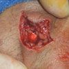
Also look for partial tendon injuries that might be missed on strength testing. Tendons run surprisingly close to the surface on the dorsum of the hand and fingers (Figure 1). Any tendon involvement requires antibiotic prophylaxis, specialty consultation, and follow-up.
Debridement. Debridement of devitalized tissue is critical to minimizing the risk of infection. Devitalized tissue can usually be identified by its dusky appearance and lack of or limited connection to the subcutaneous blood supply. However, avoid the temptation to "straighten up" wound margins when tissue is viable. Jagged wound edges aid in proper closure and minimize scarring. Unnecessary debridement increases wound tension and often leads to a wider and more noticeable scar.
STRATEGIES TO MINIMIZE INFECTION RISK
The overall rate of infection in wounds of any kind is about 3%. Factors that lower this risk include adequate debridement and irrigation, and the use of tape or staples rather than suture. "Loose" closure has been shown not to diminish risk. Factors that increase the risk of infection include comorbidities, such as heart, liver, or kidney disease and diabetes; use of epinephrine; edema; delayed presentation; crush injuries; and foreign bodies.
Consider skin closure strips for smaller wounds that are contaminated or in a location where the risk of infection is higher (eg, intra-oral wounds). Lacerations that are smaller than 1 cm often heal well without formal closure or when managed with skin closure strips alone.
Delaying-or skipping-primary closure. Closing a wound too soon may increase infection risk in certain settings. In some cases, primary closure is contraindicated. For example, puncture wounds and animal bites to the hands or feet should almost never be closed, mainly because of the increased risk of infection in such wounds.
When wounds are more than 8 hours old, the risks and benefits of primary closure must be carefully weighed. Primary closure can be considered:
- If the wound is clean.
- If it is on the head or face.
- If it can be properly irrigated.
However, delaying primary closure in wounds more than 8 hours olddecreases the risk of infection and may be the wisest course:
- If the wound is dirty.
- If it is on an extremity or the torso.
- If it is too small to irrigate properly.
- If it has devitalized tissue.
Also consider delaying primary closure for:
- Infected or heavily contaminated wounds.
- Bite wounds.
- Wounds that are more than 8 to 12 hours old (older than 24 hours for wounds on the head or face).
When primary closure is delayed, the wound is still anesthetized, explored, cleaned, and debrided as for initial primary closure. It is then packed open and re- checked in 36 to 48 hours and again after 3 to 5 days. If the wound does not appear infected at the time of the second check, it can then be sutured. Cosmetic results are similar to those achieved with early primary closure, but the risk of infection is much lower.
Antibiotic prophylaxis. In lacerations, the use of antibiotic prophylaxis is somewhat controversial. However, there is evidence to support prophylactic antibiotic use in the following settings:
- Open fractures.
- Tendon injuries.
- Animal bites to the extremities.
- Presence of lymphedema.
- Presence of a heart murmur.
You may also want to consider antibiotics in other wounds with high-risk features, especially if several of these features are present.
Tetanus booster. With clean lacerations, administer a tetanus booster if it has been more than 10 years since the last vaccination-with high-risk wounds, if it has been more than 5 years.
PREPARATION OF THE SKIN
Topical antiseptics such as povidone-iodine may be used on intact skin, but they should be kept out of the wound, or at least washed out if they do get in. Also make sure that hair is kept out of the wound: either trim the hair or use a topical antibiotic ointment to mat it down. If you trim a patient's hair, use scissors or electrical clippers; razors increase the risk of infection. (Most texts suggest avoiding shaving eyebrows because these may not grow back.) Follow preparation of the skin with a sterile prep and drape.
SELECTION OF SUTURE
Each type of suture has advantages and disadvantages. Thinner suture leaves smaller holes in the skin and is appropriate for the face, areas of cosmetic concern, and areas of low tension where strength to resist breaking is not an issue. Thicker suture is stronger and is appropriate for the extremities and areas of higher wound tension, such as near joints. Many clinicians prefer to use 6.0 or sometimes 5.0 on the face, and 4.0 in most other areas. On the sole of the foot or in areas with high tension where there is a risk of breakage, 3.0 suture can be used.
Absorbable suture is appropriate for buried stitches and for patients who may not be able to return for suture removal. Also consider using absorbable suture in uncooperative patients, such as young children, especially if conscious sedation is required to place the stitches.
Many clinicians prefer the feel of nylon to that of polypropylene; nylon also has better workability and knot security. However, polypropylene has better strength and tissue reactivity; it also may be the better choice when stitches must be placed in dark eyebrows or other areas where its blue color may help make the stitches more visible, facilitating their removal. Other features of some of the more popular types of suture are compared in the Table.
STITCHING BASICS
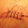
There are many ways to suture a wound. Most emergency department physicians use primarily interrupted stitches (Figure 2), while most surgeons use primarily running stitches. I prefer running stitches; their main advantage is that they save time. They also distribute tension more evenly and thus can minimize tissue strangulation. In a clean linear wound, a single long running stitch may be all that is needed.
If there is tension on a wound or if it has irregular areas, interrupted stitches may be required. Some clinicians also prefer to use interrupted stitches in infection-prone wounds: if the wound becomes infected in just one area, some of the stitches can be removed while others are left.
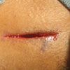
Figure 3
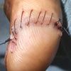
Figure 4
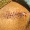
Figure 5
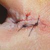
With running stitches, the first stitch and tie are the same as for a simple interrupted stitch. I generally start at one edge of the wound, although I may start in the center if there is tension. With a linear wound, I stretch the wound (to avoid translational errors) as I begin my first stitch (Figure 3). If there is tension on the wound, I "lock" the stitch after the first throw by pulling both sides of the suture to one side. This holds the first throw tight and helps prevent a loose first knot. I then cut the short end and continue suturing with the long end, making sure that the second stitch is close to the first. Each stitch should run perpendicular to the wound below the surface, leaving the visible portions of suture at a slight angle to the wound (Figure 4). The final 2 stitches, like the first 2, should also be close together. Tie the final knot using a "bite" of suture that is a loop rather than a single loose end (Figure 5). Because suturing done with running stitches relies on a single knot at each end, I make sure that my knots have at least 5 throws each.
STITCHES FOR SPECIAL SETTINGS
Other, less frequently used suturing techniques include:
- Vertical mattress stitch.
- Horizontal mattress stitch.
- Interlocking stitch.
- Subcuticular stitch.
- Buried stitches.
Mattress stitches. The vertical mattress stitch might be described as 2 stitches in 1. It is useful in settings in which you do not want to place a deep stitch because of tension on a wound.
A horizontal mattress stitch is basically 2 stitches side by side. However, in a regular interrupted stitch the suture material crosses above the wound, while in a horizontal mattress stitch, it does not (Figure 6). Use of the mattress stitch helps maintain wound eversion and may decrease suture time by halving the number of knots that need to be tied. Wound inversion may be a problem in skin that has minimal or loose supportive tissue, such as that of the back of the hand (where the horizontal mattress stitch is likely to work well).
A variation of the horizontal mattress stitch is a good choice for the pointed tip of a skin flap. Start on the non-flap area, then run the stitch sideways through the tip of the flap. Finally, finish as you would with a regular horizontal mattress stitch. This technique helps to minimize tissue strangulation in an area with compromised circulation.
Buried stitches. These can be helpful in wounds under tension and when there is potential dead space that needs to be closed. Buried stitches can also improve cosmetic results by minimizing wound tension at the epidermal level and decreasing hematoma formation. However, buried stitches are probably overused. They can increase infection risk, and they can work their way out to the surface weeks after the wound has healed. Newer, antibiotic-impregnated absorbable suture may reduce the risk of infection. Avoid sewing through adipose tissue; this causes strangulation and increases infection risk. When deep stitches are used, make sure the patient is aware of their presence.
Subcuticular stitches. Continuous subcuticular or running dermal sutures are frequently used by surgeons in conjunction with skin closure strips to obviate the need for suture removal. Subcuticular stitches are technically more difficult than other types. The stitches are placed parallel to the skin surface at the dermal-epidermal junction; backtrack slightly with each stitch to ensure correct positioning. Skin closure strips may be used on the surface to improve apposition.
Subcuticular stitches are a good choice for patients who are prone to keloid formation. However, avoid the use of absorbable suture in such patients; it may provoke more tissue reaction than nylon or polypropylene.
If non-absorbable suture is used, make sure that both ends exit the skin, and stitch so that the suture surfaces every 3 cm (this allows it to be easily cut into segments and thus facilitates removal). When absorbable suture is used, the entire closure may remain buried.
SAFE SUTURING
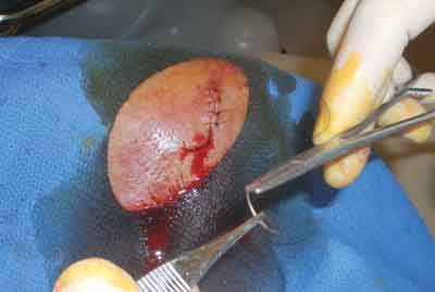
Figure 7
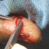
Figure 8
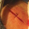
Figure 9
Always consider your own safety and comfort when you close lacerations. Use proper lighting and positioning: elevate the bed or examination table until the wound is at about the level of your elbow. Use protective goggles when injecting anesthesia. Avoid grabbing the needle with your hand; use a needle driver and regular-tip forceps to handle the needle instead. You can actually complete the entire repair without ever touching the needle with your hand (Figures 7, 8, and 9).
AFTERCARE, FOLLOW-UP, AND SUTURE REMOVAL
Once the repair is finished, I have the patient put light pressure on the wound with 1 finger for 10 minutes. This minimizes the amount of blood that oozes from the wound (oozing blood can slow healing by separating wound edges). The pressure also minimizes seepage of blood through the needle holes, which can later make suture removal more painful. In addition, a clean outer appearance looks better and may increase patient satisfaction.
After 10 minutes of light pressure, place a sterile dressing, along with a splint to prevent added wound tension when appropriate. Instruct the patient to keep the wound clean and dry (except for antibiotic ointment) for the first 48 hours to allow adequate time for an epithelial layer to form. After that, the patient may shower, and antibiotic ointment is no longer recommended for most wounds.
Wounds are usually checked within 48 hours. In high-risk wounds, a 48-hour check is obligatory.
Suture removal. The appropriate timing of suture removal depends on the location and depth of the wound, wound tension, patient age, and comorbidities. I find the following breakdown of time to suture removal useful (with allowances made for special considerations):
- After 4 to 5 days: facial wounds.
- After 15 days: extremity lacerations, wounds with tension.
- After 10 days: all other wounds.
Consider leaving sutures in for additional time in patients in whom healing may be delayed, such as elderly persons, those with compromised immune systems, and smokers. Leaving stitches in a few extra days prevents wound dehiscence from early removal, and except in facial lacerations-in which a delay in suture removal could cause "cross-track" scarring from the sutures-it has few disadvantages.
Minimizing scarring. Patients frequently ask whether they will have a scar. The appropriate answer is that all lacerations leave some form of scar, but that scarring can be minimized by appropriate sun protection. Initially, this may involve bandages and hats or clothing. Once the wound is healed, counsel patients to use sunscreen daily for at least 6 months and preferably up to a year. Advise patients in whose wounds the tension was initially great enough to hold the edges more than 5 mm apart that with time their scar is likely to widen. Also, warn patients with lacerations from blunt trauma or crush mechanisms that they have a higher risk of noticeable scarring.
References:
REFERENCES:
1.
www.ERPocketBooks.com
.
2.
Pregerson DB.
Quick Essentials Emergency Medicine.
2nd ed. Available at:
www.ERPocketBooks.com
. 2006.
