- Clinical Technology
- Adult Immunization
- Hepatology
- Pediatric Immunization
- Screening
- Psychiatry
- Allergy
- Women's Health
- Cardiology
- Pediatrics
- Dermatology
- Endocrinology
- Pain Management
- Gastroenterology
- Infectious Disease
- Obesity Medicine
- Rheumatology
- Nephrology
- Neurology
- Pulmonology
Spinal Epidural Abscess in an Obese Woman With Back Pain
An obese woman in her thirties with a history of fibromyalgia syndrome, depression, polycystic ovarian syndrome, and diabetes mellitus presents to her local emergency department with 1 week of gradually worsening midline back pain.
An obese woman in her thirties with a history of fibromyalgia syndrome (FMS), depression, polycystic ovarian syndrome, and diabetes mellitus (DM) presents to her local emergency department (ED) with 1 week of gradually worsening midline back pain. At first she thought the pain was a result of her FMS, but because it did not improve after a few days like it usually does she saw a chiropractor, which also did not help. She finally went to her regular doctor, who prescribed hydrocodone/acetaminophen and cyclobenzaprine; neither has helped her.
Over the past 24 hours, the patient has been constipated and has had difficulty with urinating. Both legs have started to feel “wobbly” and numb. The pain extends from below her neck down to her waist in the midline; it seems to move around some but usually is worst “just above her bra strap.”
The patient has no additional complaints and states that this episode definitely is not like her typical FMS attack. When asked specifically, she denies fever, abdominal pain, and vomiting.
On physical examination, the patient’s pulse is 91 beats per minute; blood pressure, 138/76 mm Hg; respirations, 22 breaths per minute, with a pulse oximeter reading of 99%; and temperature, 37.4°C (99.4°F) taken orally.
Findings from inspection of the patient’s head and neck are unremarkable, but the astute doctor, suspecting the worst, checks for meningismus. The presence of fever and back pain indicates that she has it. Her chest examination findings are completely normal. Her back and flank areas are not particularly tender and neither is her abdomen, but she is quite obese.
Findings ffrom the rest of the examination are unremarkable except for the neurological examination, which shows subjective decreased pinprick sensation in both legs and even up to the lower abdomen. The patient has normal distal leg strength with both plantar flexion and dorsiflexion of the foot, but when she undergoes a straight-leg raise test, she can keep her heels up for only 1 or 2 seconds.
A spinal MRI scan is ordered, but the radiologist refuses to call in the techs for an after-hours MRI scan without doing a CT scan first. He states that a CT scan of the spine will pick up “anything of consequence,” and if the results are negative, they can always do an MRI tomorrow “if indicated.”
A cut from the CT scan at the level of maximal pain is shown here. What do you see? What should you do next?
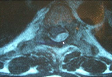
You cannot see enough on the CT scan, which shows only the vertebral body and the spinal canal, with no detail. This test is inadequate. You need the MRI scan (below), which shows a spinal epidural abscess (SEA) (white arrow). DM was this patient’s only risk factor. Her presentation, including the initial miss by her primary care physician, is fairly classic. She was taken emergently to surgery for evacuation of the abscess.

Discussion
The diagnosis of SEA, a somewhat rare condition, often is late because its presentation is indolent and the vast majority of patients who present with similar complaints have other, less serious conditions, such as musculoskeletal back pain and renal disorders.
If the diagnosis is early, surgical evacuation and antibiotics can provide a chance for a good outcome. However, delays in diagnosis and care often result in permanent neurological deficits. The patient’s future hangs in the balance, and the astute clinician who remains vigilant will be best positioned to make a “great save.”
Clues that help differentiate SEA from more routine causes of back pain are as follows:
• Pain with SEA usually develops insidiously and is most common in the thoracic area; other spinal conditions primarily affect the cervical and lumbar areas.
• Fever is a major red flag for this condition, but it may not be present, especially early in the course of disease.
• Cord tethering may be noted as a positive straight-leg raise test result or, more frequently, as meningismus, especially when the thoracic spine is involved.
• Any potential neurological complaint that involves more than one extremity is a big clue, as in this case, that the spinal cord and not just a nerve root is being affected.
• Constipation and urinary difficulty certainly may be caused by narcotic medications, such as hydrocodone, but they should be assumed to be the result of cord compression until proved otherwise.
• A post-void residual urine volume measurement and a rectal examination can help distinguish a neurological condition from a medication adverse effect, but if doubt still exists, neuroimaging is required.
• Risk factors for SEA include injection drug abuse; any invasive procedure; immunosuppression, such as with DM; and a concomitant bacterial infection (Table).
The diagnostic study of choice for SEA is spine MRI; the thoracic spine should be included even when neurological findings appear to come from the cervical or lumbar area. If MRI is unavailable, CT myelography is an acceptable alternative.
A CT scan with contrast may be considered as a screening test because results often are available more rapidly. However, it should not be used to exclude the diagnosis of SEA because false negatives may occur for a variety of reasons, including obesity, as in this case.
SEA is very unlikely to occur in the setting of a normal erythrocyte sedimentation rate.
Treatment of patients with SEA involves emergency surgery as well as the use of antibiotics. Surgery should prevent progression of neurological damage; in the long run, however, the patient often is left with the same degree of neurological deficit present at the time of surgery. Therefore, early diagnosis and rapid surgical decompression are essential for reaching the best possible outcome (see Table).
The patient is this case did well and had a good recovery, although she was left with some mild weakness in her legs. Key aspects of her ED care were early recognition that her current symptoms were significantly different from her typical back pain episodes; appropriate concern about the presence of bilateral leg symptoms; the presence of DM as an important risk factor for SEA; and early recognition of a low-grade fever, meningismus, and mild leg weakness on physical examination.
The radiology department’s refusal to expedite the MRI scan was less than ideal. However, the CT results were available promptly so that minimal time was lost.
Delayed diagnosis in 50% of patients because initially insidious; progression later becomes rapid
1. Back pain that may start as mild and often is thoracic, fever
2. Nerve root pain (sciatica or cervical)
3. Motor and sensory findings
4. Paralysis from cord compression
Early: Spinal tenderness, positive straight-leg raise test result, meningismus, fever in 50% of patients
Late: Sensory level (a vertebral level below which sensation is absent), weakness, urinary retention, decreased rectal tone
Staphylococcus, Escherichia coli, Pseudomonas; concomitant discitis, osteomyelitis, or endocarditis common
Central line (central venous catheter), injection drug abuse, recent procedure, acupuncture
Depressed immunity: diabetes mellitus, cancer, corticosteroid, alcohol, kidney or liver failure, pregnancy
Distant infection (UTI, pneumonia, neck furuncle), spinal fracture
Best: MRI with contrast or CT myelography
Alternatives: CT with contrast (50% sensitive), ESR > 20 mm/h (reportedly 98% sensitive)
Psoas or deep neck abscess, meningitis, herniated disc, UTI, discitis, osteomyelitis
Surgery stat with spine surgeon (spine orthopedist or neurosurgeon)
Antibiotics: ceftriaxone 1 g IV + clindamycin 300 mg IV + vancomycin 1 g IV or alternative
UTI, urinary tract infection; ESR, erythrocyte sedimentation rate. Copyright Emresource.org.
