- Clinical Technology
- Adult Immunization
- Hepatology
- Pediatric Immunization
- Screening
- Psychiatry
- Allergy
- Women's Health
- Cardiology
- Pediatrics
- Dermatology
- Endocrinology
- Pain Management
- Gastroenterology
- Infectious Disease
- Obesity Medicine
- Rheumatology
- Nephrology
- Neurology
- Pulmonology
Something Wrong on the Face of an Old Man
A 76-year-old man is seen because of redness below the right eye. Has long-standing “lazy eye” on the left, which is chronically deviated outward. Has lived in nursing home for some years due to self-care deficit from memory loss. No recent eye surgery, conjunctivitis, sinus infection, or periocular trauma.
This article was originally presented as an independent educational activity under the direction of CME LLC. The ability to receive CME credits has expired. The article is now presented here for your reference. CME LLC is no longer responsible for the presentation of the article.
HISTORY

A 76-year-old man is seen because of redness below the right eye. Has long-standing “lazy eye” on the left, which is chronically deviated outward. Has lived in nursing home for some years due to self-care deficit from memory loss. No recent eye surgery, conjunctivitis, sinus infection, or periocular trauma.
PHYSICAL EXAMINATION
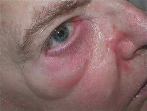
Man who does not effectively communicate how much of his moderate discomfiture is psychological versus physical. Temperature, 37.3°C (99.2°F); heart rate, 84 bpm and regular; BP, 126/84 mm Hg; respirations, 20 per minute and not labored. Face as shown. Visual function seems preserved within limits of his replies. Full ocular range of motion. Each pupil constricts on direct illumination and consensually on shining flashlight beam into opposite eye.
What’s Your Diagnosis?
ANSWER: PRESEPTAL CELLULITIS WITH EARLY FACIAL CELLULITIS
This man’s localized redness about the right orbit, centered on the lower lid, permits a diagnosis of preseptal cellulitis,1-5 a more common and milder bacterial infection than the frank orbital cellulitis that affects more posterior structures when infection breaches the orbital septum.
In overt orbital cellulitis, the patient often has abrupt onset of substantial fever and prostration, in contrast to the status of this patient. Examination in the more severe condition often shows proptosis when inflammatory exudate presses the globe forward, and typically downward as well. As might be predicted given involvement of deeper ocular and orbital tissue, the ocular range of motion can be impaired as can visual acuity and even pupillary light reactions; painful diplopia and pain on eye movement announce this is far worse than simpler causes of red eyes.

Figure 1 – Closeup view of right eye and orbit shows that the periocular swelling and erythema are confined strictly to the inferior area, sparing the upper lid. The conjunctiva is free of vascular dilation or erythema, and of chemosis. There is no exudate residue on the lashes. On this closer view it is also especially clear that no proptosis is present.
Our view of this patient’s visible eye tissues aids in recognizing that he has the milder preseptal cellulitis: with his light-colored irides, one readily discerns that the pupil on the affected right side is the same size as on the left or just a tad smaller (Figures 1and 2); therefore, pupilloconstrictor fibers of the third cranial nerve are unaffected; ocular range of motion testing showed other functions of III, IV, and VI intact; and his replies about objects we asked him to identify with each eye individually- the other being passively covered by our hands-confirmed that at least grossly, acuity was unaffected. There is no conjunctivitis or keratopathy. The eye is not protuberant, so he has no proptosis. Preseptal cellulitis may not come to our minds because of sparing of the upper lid: a red, swollen upper lid is the most recognized visual clue. However, once we identify the case as some subtype of orbital infection, there is no doubt where this falls on the scale of severity: mild.

Figure 2 – Left eye provides perfect image of baseline. Because the eyes are turned to the left, the weakness of the left medial rectus is hidden and the eyes appear conjugate though pinpoint light reflections still vary from right eye (iris) to left (pupil). Fine telangiectases on nose, cheek, and left ala nasi hint that right-sided nasal erythema may be completely unrelated to the cellulitis.
In trying to make sense of changes away from the orbit, the cutaneous redness is patchy and variable: the “satellite” zone abutting the right ala nasi appears more salmon-colored and shinier (see Figure 1); one can’t be certain if it is related to the orbital swelling or perhaps from an entirely unrelated cause such as a preexistent rosacea (see Figure 2). Drooping of the lower cheek on the right hints that there could be swelling there as well, but the redness near there is so slight that we conclude weakly, “Maybe a touch of facial cellulitis as well.”
GRAVE COMPLICATIONS OF ORBITAL CELLULITIS
Dreaded complications occur in an important subset of cases of orbital cellulitis: meningitis, brain abscess, and cavernous sinus thrombosis. If one recalls the contents of the cavernous sinus as “III, IV, V-1, V-2, VI, internal carotid artery”-as my old friend and laboratory partner and I do, in cadence, from the teaching of our anatomy laboratory instructor decades ago-one knows that the signs to look for, besides prostration, delirium, and fever, will be pupillodilation; loss of ocular range of motion overall from III and IV, and of lateral deviation of the eye ipsilateral to the lesion due to involvement of the lateral rectus-abducens complex; and loss of upper- and mid-facial sensation. Blindness can ensue, and so, with carotid involvement, can stroke and death. Given the stakes, ophthalmological consultation will be mandatory in any case of frank orbital cellulitis, and hospitalization will almost surely follow; in contrast, a confident diagnosis of preseptal cellulitis mandates oral antibiotics and close follow-up. Ongoing research to classify severity and to guide treatment considers the biological state of the host, and the reliability of early return if improvement is not occurring promptly.3-5
Sinus infection, especially of the ethmoid, underlies many cases of frank orbital cellulitis; other cases follow eye surgery or penetrating ocular trauma. Simpler problems of the lids or external eye, such as hordeolum (stye) or insect bites, are more commonly antecedents of preseptal cellulitis. Both eye infections occur more often in children, adolescents, and young adults.
DIVERSE CAUSES OF PRESEPTAL CELLULITIS
Although conventional Gram-positive cocci account for many cases of preseptal cellulitis, a polymicrobial etiology is common in orbital cellulitis and often includes anaerobes, typically in a synergistic relationship with other species.
The list of rare microbial causes of preseptal cellulitis goes on and on, from vaccinia due to live-viral smallpox vaccine6 up the ladder of complexity to Chlamydia7- more familiar as a cause of ordinary conjunctivitis-to Bacillus anthracis as part of the spectrum of cutaneous anthrax,8 to gonococcus,9 which is again much more familiar as a pure conjunctival pathogen in ophthalmia neonatorum. Mycobacterium tuberculosis has caused preseptal cellulitis,10 and so have ringworm fungi.11 Even fly infestation has been demonstrated12; we know that helminthic infection with prominent eosinophil reaction causes orbital muscle involvement in trichinosis.
Clinical manifestations of preseptal cellulitis have been characterized, but new variants arise: necrosis of lids in streptococcal infection,13 reminiscent of the “flesh-eating bacteria” of necrotizing fasciitis. Community- acquired methicillin-resistant Staphylococcus aureus (CAMRSA) has, predictably, caused recurrent preseptal cellulitis in an otherwise healthy athlete with an under-lying eczema. That particular individual had leukocytosis but no fever.14
If the list of causes were not long enough, preseptal cellulitis has also been produced by habitual picking and self-mutilation,15,16 both with and without the intermediary of bacterial infection; and by the local effects of cocaine abuse.17
MULTIPLE EXPLANATIONS, MULTIPLE THEORIES, MULTIPLE DISTRACTING COMORBIDITIES
The present patient illustrates quite dramatically the challenge of diagnosis in elderly persons whose background conditions distract us; and his replies on tests that require cooperation must be viewed with circumspection.
Specifically, one forms an impression that he has hypertelorism and even perhaps proptosis of the left eye. One wonders if the impression is misleading and due to his dysconjugate gaze. The gaze deviation is underscored, in addition to the asymmetry of eye position, by discordant light reflections, from the center of the pupil on the right; but from the medial iris, well away from mid-pupil, on the left. The divergent gaze is attributable to stable exotropia (out-turned eye) on the left, and thus cannot support any reasoning about oculomotor dysfunction of the eye on the side with the redness.
The right eye is not proptotic; one cannot make a case for ptosis of its upper lid even though the height of skin exposed above it and beneath the brow is far wider than on the left: clearly that enhanced distance (“wider stripe of skin”) results from the right eyebrow’s riding higher than the left. Forehead wrinkling is intact bilaterally, so there can’t be a peripheral seventh (facial) nerve palsy, notwithstanding that the right angle of the jaw seems to sag, and the right nasolabial fold is less fully defined than its counterpart on the left. The right corner of the mouth dips: at most we have a subtle right central facial palsy, but this can’t be linked to an abnormality of superficial layers of the eye, eg, exposure keratopathy (see Figure 1). Once again, major abnormalities are present but irrelevant to, and confounding of, diagnosis of the acute problem on the right.
Fortunately, one can often, as here, separate diagnostic features from red herrings. Knowledge of the patient from beforehand is an immense help in this regard- yet another argument for continuity of care, for no chart provides the level of awareness of baseline that multiple prior visits can do. And should we not welcome a diagnostic challenge? If all our patients had a single problem or a classic presentation, we’d find clinical diagnosis too easy and then boring, something of which there is no danger at present.
References:
REFERENCES:
1
. Kanski JJ.
Clinical Ophthalmology: a Systematic Approach.
3rd ed. Oxford andLondon: Butterworth Heinemann; 1994:38-39.
2
. Hasanee K, Sharma S. Ophthaproblem. Orbital cellulitis.
Can Fam Physician
.2004;50:359, 365, 367.
3
. Chaudhry IA, Shamsi FA, Elzaridi E, et al. Inpatient preseptal cellulitis: experiencefrom a tertiary eye care centre.
Br J Ophthalmol
. 2008;92:1337-1341.
4
. Liu IT, Kao SC, Wang AG, et al. Preseptal and orbital cellulitis: a 10-year reviewof hospitalized patients.
J Chin Med Assoc
. 2006;69:415-422.
5
. Vu BL, Dick PT, Levin AV, Pirie J. Development of a clinical severity score forpreseptal cellulitis in children.
Pediatr Emerg Care
. 2003;19:302-307.
6
. Hu G, Wang MJ, Miller MJ, et al. Ocular vaccinia following exposure to asmallpox vaccinee.
Am J Ophthalmol
. 2004;137:554-556.
7
. Drummond SR, Diaper CJ. Chlamydial conjunctivitis presenting as preseptalcellulitis.
Head Face Med
. 2007;3:16-18.
8
. Artac H, Silahli M, Keles S, et al. A rare cause of preseptal cellulitis: anthrax.
Pediatr Dermatol.
2007;24:330-331.
9
. Green JA, Lim J, Barkham T.
Neisseria gonorrhoeae
: a rare cause of preseptalcellulitis?
Int J STD AIDS
. 2006;17:137-138.
10
. Raina UK, Jain S, Monga S, et al. Tubercular preseptal cellulitis in children:a presenting feature of underlying systemic tuberculosis.
Ophthalmology
. 2004;111:291-296.
11
. Rajalekshmi PS, Evans SL, Morton CE, et al. Ringworm causing childhoodpreseptal cellulitis.
Ophthal Plast Reconstr Surg
. 2003;19:244-246.
12
. Jun BK, Shin JC, Woog JJ. Palpebral myiasis.
Korean J Ophthalmol.
1999;13:138-140.
13
. Stone L, Codere F, Ma SA. Streptococcal lid necrosis in previously healthychildren.
Can J Ophthalmol
. 1991;26:386-390.
14
. Charalampidou S, Connell P, Fennell J, et al. Preseptal cellulitis caused bycommunity acquired methicillin resistant Staphylococcus aureus (CAMRSA).
Br J Ophthalmol
. 2007;91:1723-1724.
15
. Ugurlu S, Bartley GB, Otley CC, Baratz KH. Factitious disease of periocularand facial skin.
Am J Ophthalmol.
1999;127:196-201.
16
. Trager MJ, Hwang TN, McCulley TJ. Delusions of parasitosis of the eyelids.
Ophthal Plast Reconstr Surg
. 2008;24:317-319.
17
. Underdahl JP, Chiou AG. Preseptal cellulitis and orbital wall destructionsecondary to nasal cocaine abuse.
Am J Ophthalmol.
1998;125:266-268.

