- Clinical Technology
- Adult Immunization
- Hepatology
- Pediatric Immunization
- Screening
- Psychiatry
- Allergy
- Women's Health
- Cardiology
- Pediatrics
- Dermatology
- Endocrinology
- Pain Management
- Gastroenterology
- Infectious Disease
- Obesity Medicine
- Rheumatology
- Nephrology
- Neurology
- Pulmonology
Skin Disorders in Older Adults: Papulosquamous and Bullous Diseases, Part 2
Here: a succinct review of the diagnosis and treatment of pemphigus vulgaris, dermatomyositis, cicatricial pemphigoid, linear IgA bullous disease, coma bullae, and stasis bullae.
ABSTRACT: Certain papulosquamous and bullous diseases are more common in older adults than in younger persons. For example, the mean age of onset of pemphigus vulgaris is about 50 to 60 years. Because this disease can be life-threatening, prompt diagnosis is crucial. In elderly persons, dermatomyositis often signals underlying malignancy. Cicatricial pemphigoid and linear IgA bullous disease are rare subepidermal blistering diseases that occur more frequently in older adults. Coma bullae are typically seen in elderly patients who have fallen and lain unconscious for hours or days. Stasis bullae can develop in patients who have limited or no mobility. These lesions result from chronic edema caused by hypoalbuminemia, venous thrombosis, congestive heart failure, or hepatic and renal failure.
Key words: pemphigus vulgaris, dermatomyositis, cicatricial pemphigoid, linear IgA bullous disease, coma bullae, stasis bullae
Although inflammatory dermatoses are generally less prevalent among elderly persons than among younger adults, some of these dermatoses-particularly vesiculobullous eruptions-are more common in patients over age 60. In this second part of my 2-part series on papulosquamous and bullous cutaneous diseases in older adults, I review the diagnosis and treatment of pemphigus vulgaris, dermatomyositis, cicatricial pemphigoid, linear IgA bullous disease, coma bullae, and stasis bullae. In part 1 (CONSULTANT, March 2011, page 168), I discussed psoriasis, bullous pemphigoid, and Grover disease.
PEMPHIGUS VULGARISClinical features. Pemphigus vulgaris belongs to the group of systemic autoimmune blistering diseases of the skin and mucous membranes. The mean age of onset is about 50 to 60 years. Because pemphigus can be life-threatening, prompt diagnosis is crucial.
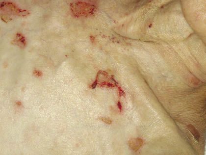
Figure 1 – The flaccid blisters of pemphigus vulgaris rupture easily and leave painful erosions, as seen in this elderly woman.
In patients with pemphigus vulgaris, flaccid blisters develop on the skin; these rupture easily, leaving painful denuded surfaces (Figure 1) that can sometimes be crusted (Figure 2) and are often geometric in shape. Bullae and erosions typically manifest on the upper trunk; head; neck; and intertriginous areas, such as the axillae (Figure 3) and groin. The disease usually begins in the oral cavity. It may be localized, or it may become generalized and involve 20% to 50% of the skin in severe disease. Involvement of ocular, nasal, pharyngeal, laryngeal, esophageal, vaginal, penile, and anal mucosa typically occurs in severe disease. Healing usually occurs without scarring and is often accompanied by postinflammatory hyperpigmentation.
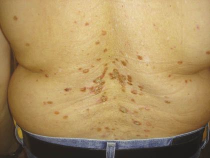
Figure 2 – Pemphigus vulgaris manifests in this man as crusted plaques.
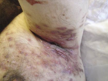
Figure 3 – Extensive axillary involvement is evident in this man with pemphigus vulgaris.
Treatment. Corticosteroids are the mainstay of treatment. Immunosuppressants, such as mycophenolate mofetil and azathioprine, can be used as corticosteroid-sparing agents. Rituximab and intravenous immunoglobulin have also been effective in patients with pemphigus vulgaris.1
DERMATOMYOSITIS
Clinical features. This systemic disorder manifests with inflammatory myopathy and cutaneous eruptions. The typical age of onset is in the 50s and 60s. Note that in elderly persons, dermatomyositis is often associated with underlying malignancy. In adults who are older than 50 years, the most common associated malignancies are those of the breast, colon, lung, ovary, stomach, and uterus. Ovarian cancer has the strongest association with paraneoplastic dermatomyositis, which affects women more than men, in a ratio of 2:1.2
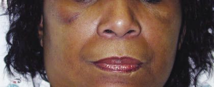
Figure 4 – The characteristic heliotrope rash of dermatomyositis is seen in this woman.
Dermatomyositis has a variety of cutaneous manifestations. Distinctive red, blue, or purple patches and plaques are often present. The characteristic heliotrope rash is an eruption over the eyelids and periorbital area; the color resembles that of the heliotrope flower (Figure 4). Gottron papules are another characteristic feature of dermatomyositis. These are flat-topped violaceous papules that vary in degree of atrophy; they occur on the dorsa of the knuckles (Figure 5)-sparing the interarticular areas, which lupus commonly affects-and on extensor surfaces of the skin on the joints of the hands, knees, and elbows.
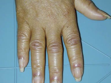
Figure 5 – Gottron papules occurred on the dorsa of the knuckles of this patient with dermatomyositis.
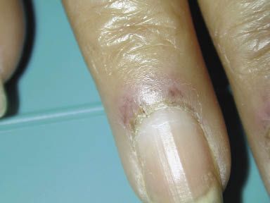
Figure 6 – This close-up of the finger of a woman with dermatomyositis shows the characteristic manifestations of periungual erythema, ragged cuticles, and telangiectases.
Dermatomyositis can present with erythema of the face, neck, upper trunk, knuckles, elbows, and knees. Poikiloderma (atrophic skin with dilated blood vessels and brown postinflammatory pigmentation) may also be noted in a photosensitive distribution. Other manifestations of dermatomyositis include periungual erythema, ragged cuticles, telangiectases, thrombosis of capillary loops, and infarctions on the fingers (Figure 6). Erosions (Figure 7), ulcerations, and desquamation can occur. Patients may report a scaly scalp and thinning of the hair.
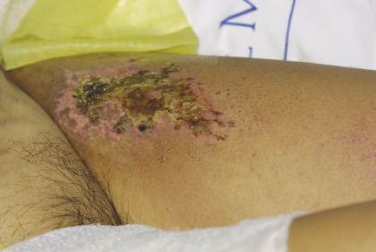
Figure 7 – Erosions and ulcerations can occur in patients with dermatomyositis.
Diagnosis. Diagnostic criteria consist of proximal muscle weakness, elevated serum muscle enzyme levels, diagnostic muscle biopsy, and characteristic changes on electromyography.3 The evaluation of patients with suspected dermatomyositis should include skin biopsy and measurement of creatine kinase, aldolase, aspartate aminotransferase, lactate dehydrogenase, glutamic oxaloacetic transaminase, antinuclear antibody, anti-Mi-1, anti–signal recognition protein, anti-Ku, and urinary creatinine. Pulmonary function studies and electrocardiography may also be performed. Esophageal manometry may be appropriate for certain patients. Other tests include MRI, chest radiography (to detect interstitial fibrosis), and barium swallow studies.
Treatment. I typically refer patients with dermatomyositis to a rheumatologist for treatment.
CICATRICIAL PEMPHIGOID
Clinical features. Also known as “mucous membrane pemphigoid,” this rare, chronic subepidermal blistering disease has a mean age of onset of 73 years. Women are affected twice as frequently as men. There is no racial predilection. The reported incidence varies from 0.87 to 1.16 cases per million persons.
Cicatricial pemphigoid usually affects the mucosa and occasionally the skin. It often remains localized to the oral mucosa, and the most common presentation is desquamative gingivitis. Other areas of the oral cavity can also be involved, including the buccal mucosa, palate, tongue, and lips.
In some patients, the predominant involvement is conjunctival, and the disease is then sometimes termed “ocular cicatricial pemphigoid.” Patients with ocular involvement characteristically have symptoms of conjunctivitis, including irritation, burning, increased lacrimation, photophobia, and dryness of the eyes. These symptoms are initially unilateral but can become bilateral within a period of 2 years.
Cicatricial pemphigoid can also involve the nasopharyngeal, laryngeal, anogenital, vaginal, penile, and esophageal mucosae. Cutaneous lesions can present on the face, neck, scalp, axillae, trunk, and extremities.
Bullae and erosions heal with scarring in most but not all instances. This scarring is often responsible for the major morbidity and irreversible sequelae associated with cicatricial pemphigoid. The scarring of the conjunctiva can progress to blindness if it is not treated early.
Treatment. In some patients with cicatricial pemphigoid who have limited oral involvement, topical corticosteroids and, most recently, tacrolimus have been reported to control the disease.4 Most patients who have progressive disease require an aggressive treatment regimen. High doses of systemic corticosteroids and dapsone are most often used as first-line agents in this setting.
LINEAR IgA BULLOUS DISEASE
Clinical features. The reported incidence of this subepidermal blistering disease is less than 0.5 per million in western Europe.5 The mean age of onset is 60 years, and the disease is more common in women than in men. Linear IgA bullous disease has been associated with certain systemic diseases, including chronic lymphocytic leukemia, ulcerative colitis, and dermatomyositis. Patients can present with both mucosal and cutaneous involvement. Cutaneous manifestations consist of vesicles along with urticarial plaques in an arcuate pattern, distributed over the trunk and extremities.
Treatment. The mainstay treatment of linear IgA bullous disease is dapsone. An initial response to dapsone can be seen as early as within 72 hours of initiating treatment. The doses of dapsone used are similar to those used in other autoimmune blistering diseases.
COMA BULLAE
Clinical features. Coma bullae occur on body areas to which pressure has been applied. They arise as tense blisters without much erythema or as urticarial plaques, which evolve over 48 hours in most cases to blisters (Figure 8). The lesions are limited to cutaneous involvement and are most commonly observed over bony eminences.
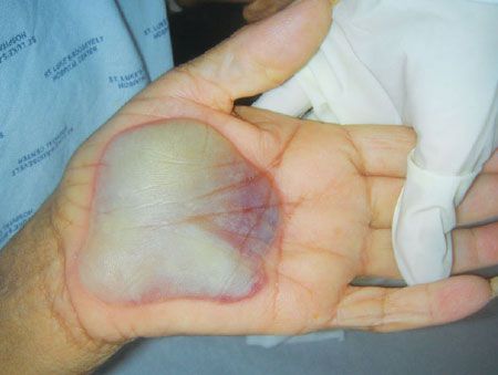
Figure 8 – A coma bulla is shown here on the palm of an elderly woman.
Patients who are receiving dialysis for chronic renal failure and those who have diabetic ketoacidosis, hypercalcemia from hyperparathyroidism, and neurological conditions are at increased risk for coma bullae. Typically, these lesions are seen in elderly patients who have been admitted to the hospital after falling to the floor and lying unconscious for hours if not days. Use of opiates, tricyclic antidepressants, antipsychotics, glutethimide, and methaqualone has been linked to coma bullae.6
Diagnosis. The biopsy of a coma bulla demonstrates the presence of a subepidermal bulla and eccrine gland necrosis. The results of direct immunofluorescence can be negative or at times can demonstrate the deposition of IgM and C3 in the dermal vessels.
Treatment. This involves the removal of pressure to the affected areas and prevention of the conditions that led to immobility. The early recognition and treatment of these lesions can prevent further consequences, such as penetration of lesions into muscle and adipose tissue. Healing occurs without scarring over a period of 2 to 4 weeks.
STASIS BULLAE
Clinical features. Stasis bullae can develop in patients who have limited or no mobility. These lesions result from chronic edema caused by hypoalbuminemia, venous thrombosis, congestive heart failure, or hepatic and renal failure. They are most commonly found on the arms and legs (Figure 9).
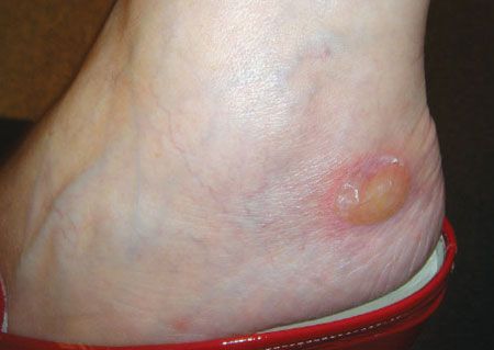
Figure 9 – This stasis bulla developed in an elderly woman with chronic edema.
Treatment. This involves correction of the underlying disease state and elevation of the extremity, compression stockings, diuretics, and prevention of infection. The bullae should not be pricked because the fluid they contain is the optimal medium for skin regrowth.
References:
REFERENCES:
1. Ahmed AR, Spigelman Z, Cavacini LA, Posner MR. Treatment of pemphigus vulgaris with rituximab and intravenous immune globulin. N Engl J Med. 2006;355:1772-1779.
2. Fardet L, Dupuy A, Gain M, et al. Factors associated with underlying malignancy in retrospective cohort of 121 patients with dermatomyositis. Medicine (Baltimore). 2009;88:91-97.
3. Dalakas MC, Hohlfeld R. Polymyositis and dermatomyositis. Lancet. 2003;362:971-982.
4. Günther C, Wozel G, Meurer M, Pfeiffer C. Topical tacrolimus treatment for cicatricial pemphigoid. J Am Acad Dermatol. 2004;50:325-326.
5. Bernard P, Vaillant L, Labeille B, et al. Incidence and distribution of subepidermal autoimmune bullous skin disease in three French regions. Bullous Diseases French Study Group. Arch Dermatol. 1995;131:48-52.
6. Waring WS, Sandilands EA. Coma blisters. Clin Toxicol (Phila). 2007;45:808-809.
