- Clinical Technology
- Adult Immunization
- Hepatology
- Pediatric Immunization
- Screening
- Psychiatry
- Allergy
- Women's Health
- Cardiology
- Pediatrics
- Dermatology
- Endocrinology
- Pain Management
- Gastroenterology
- Infectious Disease
- Obesity Medicine
- Rheumatology
- Nephrology
- Neurology
- Pulmonology
Ringworm-or Mimic?
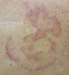
Which of the rashes pictured here is tinea corporis?
Which of the rashes pictured here is tinea corporis?


A. A large, intensely pruritic rash on the back of a man with diabetes.
B. Widespread, asymptomatic lesions on a man's trunk.
C. Recurrent lesions on a woman's legs.
D. Numerous nonpruritic lesions on a 9-year-old boy.
(Answers on next page.)
Tinea Corporis
A 68-year-old man complained of intense itching on his back of about 3 months' duration. He had adult-onset type 2 diabetes mellitus, which was poorly controlled because of lack of compliance with his dietary and medical regimen.

An erythematous, scaly rash with a slightly raised border covered most of the patient's back. Results of a potassium hydroxide preparation were positive for hyphae and confirmed the clinical diagnosis of tinea corporis.
Although topical therapy might have been successful, the patient lived alone and could not reach the entire affected skin surface; therefore, he was treated with oral terbinafine, 250 mg/d. A clinical and mycologic cure was achieved within 2 weeks.
(Case and photograph courtesy of Ted Rosen, MD.)
Cutaneous T-cell Lymphoma
For 6 months, a 63-year-old man had had a widespread but asymptomatic skin eruption. His medical history was unremarkable.


Numerous annular and serpiginous, slightly scaly lesions were present on the patient's trunk. Dermatophytosis was strongly suspected, but the results of a potassium hydroxide preparation and fungal culture were negative.
Because the rash did not respond to empiric treatment with terbinafine cream, 2 skin biopsy specimens were obtained from different sites. Both specimens revealed dermal collections of atypical lymphocytes along with exocytosis of these same cells into the epidermis ("Pautrier microabscess"). These findings are pathognomonic for cutaneous T-cell lymphoma (mycosis fungoides). This cutaneous lymphoma can present with annular lesions that precisely mimic tinea infection.
The patient is being treated with topical application of ultrapotent corticosteroids and psoralen-UV-A phototherapy.
(Case and photographs courtesy of Ted Rosen, MD.)
Nummular Eczema
Multiple, uniform, scaly, round lesions-some 4 cm in diameter-first appeared on the legs of a 61-year-old woman about 10 years earlier and recurred every winter, despite application of a number of topical antifungal medications for presumed ringworm. The rash involved only the legs and was pruritic. The patient took long, hot showers, sometimes 2 a day, and used scented deodorant soap. Results of a potassium hydroxide preparation of a scraping from a leg lesion were negative for hyphae.


Exposure to cold weather, frequent bathing, and use of harsh soaps are well-known triggers of nummular eczema. This common disorder affects the extremities of middle-aged adults and worsens in low-humidity environments, especially in northern climates. The extremities are thought to be affected preferentially because of their relative paucity of sebaceous glands, which produce even less sebum as one grows older. Although the lesions may be associated with pruritus, they are not invariably pruritic.
Nummular eczema is often mistaken for a fungal infection. However, the legs are an unusual site for ringworm, and this patient had no contact with a source of fungal infection (eg, a cat or other animal). Moreover, scale is present only at the leading edge of tinea corporis lesions.
In addition to tinea, the differential diagnosis includes psoriasis, Bowen disease, and mycosis fungoides. Psoriasis usually involves other areas, such as the elbows and knees; patients often have a history of psoriatic eruptions. Bowen disease typically appears on sun-damaged skin. The lesions of mycosis fungoides may appear as scaly plaques, but they are fixed; there is no history of waxing and waning.1
Treatment of nummular eczema involves adequate skin moisturizing and avoidance of triggers, such as frequent bathing and irritating soaps, that can worsen dry skin. Application of mid- to high-potency topical corticosteroid ointments may hasten resolution. Failure of this treatment indicates the need for a biopsy.
(Case and photographs courtesy of Joe Monroe, MPAS, PA-C.)
Atypical Granuloma Annulare
A healthy 9-year-old boy presented with round, pink, slightly raised, nonpruritic lesions on the legs of several days' duration. The lesions had arisen as papules that resembled mosquito bites and had gradually enlarged. Cellulitis was suspected, and oral cephalexin was prescribed.


At follow-up 4 days later, the lesions had multiplied and enlarged to about 2 cm; they lacked a central clearing. Daily application of ketoconazole cream was prescribed; the therapy was discontinued when the results of a potassium hydroxide preparation and fungus culture revealed no hyphae.
Five days later, new lesions, with no elevation or scaling at the periphery, had developed on the left lateral trunk and right upper arm. An incision biopsy of a lesion on the left anterior thigh revealed an increased number of lymphocytes and histiocytes arranged around dermal vessels and between collagen bundles. Increased mucin deposition was visible with hematoxylin and eosin staining and was confirmed with colloidal iron staining. These findings supported the diagnosis of granuloma annulare.
This patient exhibits an atypical clinical presentation. Typically, localized granuloma annulare presents as rings with clear centers and elevated borders of continuous papules or nodules. This patient's lesions more closely resembled macules with small papules.
The localized form of granuloma annulare usually occurs in adults, most commonly in young women, and often affects the lateral or dorsal aspects of the hands and feet and extensor aspects of the arms and legs. The lesions range from 0.5 cm to 5 cm. The course varies from months to years. Involution--usually without scarring--is typical; however, 40% of cases recur.1 Familial occurrence in siblings, twins, and successive generations is uncommon.2
Possible triggers for granuloma annulare include insect bites, trauma, viral warts, erythema multiforme, and sunlight. Lesions also may develop in the scar of herpes zoster and tuberculin skin test sites.
Granuloma annulare is associated with sarcoidosis, Alagille syndrome, autoimmune thyroiditis, hepatitis C, waxing-induced pseudofolliculitis, tattoos, granulomatosis my- cosis fungoides, monoclonal gam-mopathy, myelodysplastic syndrome, Hodgkin and non-Hodgkin lymphoma, and metastatic adenocarcinoma.3 The clinical presentation may be atypical in patients with these diseases.
Many cases of granuloma annulare are mistaken for tinea corporis and other skin infections. When the diagnosis is uncertain, obtain a biopsy. Histologic findings show collagen degeneration similar to that of necrobiosis lipoidica, which may be difficult to distinguish from granuloma annulare clinically as well as histologically.
Most lesions are asymptomatic and require no treatment. If the location of a lesion necessitates treatment, intralesional injection of triamcinolone, clobetasol lotion under occlusion, or cryotherapy can be used. Disseminated lesions may respond to dapsone, isotretinoin, etretinate, hydroxychloroquine, cyclosporine, niacinamide, psoralen-UV-A, vitamin E, or zileuton.4
Based on the biopsy results, no further treatment was given. The lesions spontaneously resolved about 2 months after onset; at 1 year follow-up, they had not recurred.
(Case and photographs courtesy of Robert P. Blereau, MD.)
References:
Nummular Eczema
REFERENCE:
1.
Monroe J. Nummular eczema.
Consultant.
2005;45:274.
Atypical Granuloma AnnulareREFERENCES:1. Wells RS, Smith MA. The natural history of granuloma annulare. Br J Dermatol. 1963;75:199-205.
2. Friedman SJ, Winkelmann RK. Familial granuloma annulare: report of two cases and review of the literature. J Am Acad Dermatol. 1987;16(3, pt 1): 600-605.
3. Weedon D. Skin Pathology. 2nd ed. London: Churchill Livingstone; 2002:200.
4. Habif TP. Clinical Dermatology: A Color Guide to Diagnosis and Therapy. 3rd ed. St Louis: Mosby; 1996:899.
