- Clinical Technology
- Adult Immunization
- Hepatology
- Pediatric Immunization
- Screening
- Psychiatry
- Allergy
- Women's Health
- Cardiology
- Pediatrics
- Dermatology
- Endocrinology
- Pain Management
- Gastroenterology
- Infectious Disease
- Obesity Medicine
- Rheumatology
- Nephrology
- Neurology
- Pulmonology
Rheumatoid Arthritis: Clues to Early Diagnosis
Primary care physicians are usually the first to see patients with joint pain; consequently they represent the "front line" of RA care. This fact-coupled with the projection that the number of rheumatologists is expected to decline by 20% during the next 2 to 3 decades-underscores the pivotal role that primary care clinicians are now expected to play in the early diagnosis of RA.
A 38-year-old African American woman sought medical care because of a 1-month history of painful, swollen hands. The patient, a hospital nurse, thought at first that her symptoms might be job-related, but she became concerned when the pain persisted and was frequently accompanied by morning stiffness that lasted some mornings for about half an hour. She tried acetaminophen and ibuprofen, which helped only slightly. She denied recent infections and other joint pain but complained of fatigue of recent onset. She also denied rash, alopecia, fever, chills, sweats, chest pain, cough, shortness of breath, photophobia, headaches, weakness, and back pain. Her symptoms interfered with a number of her daily activities. She has had a long monogamous relationship with her husband.
Physical examination revealed slight tenderness and mild swelling over the proximal interphalangeal (PIP) and metacarpophalangeal (MCP) joints bilaterally but no erythema or warmth. Mild bilateral tenderness of the metatarsophalangeal (MTP) joints was also noted. All the other joints were normal. Inflammatory polyarthritis was a diagnostic consideration. Naproxen was prescribed, radiographic and laboratory studies were ordered, and a follow-up visit was scheduled.
At follow-up 2 weeks later, the patient still had joint pain and swelling, and her fatigue had worsened, although she had difficulty sleeping because of the pain. Plain radiographs of the hands and feet revealed periarticular soft tissue swelling around 2 of the MCP joints. Laboratory results were normal except for an erythrocyte sedimentation rate (ESR) of 46 mm/h. Results of tests for rheumatoid factor (RF), antinuclear antibodies, and parvovirus antibodies were negative. The patient was referred to a rheumatologist for further evaluation; however, the next available appointment was not for 4 months.
By the time the patient saw the rheumatologist, she was in considerable pain. Swelling, redness, and warmth were present bilaterally in the PIP, MCP, and MTP joints as well as in both wrists. Morning stiffness had increased to 2 hours. Plain radiographs revealed soft tissue swelling of both hands, accompanied by periarticular osteopenia in some MCP joints. Laboratory studies revealed positive RF and cyclic citrullinated peptide (anti-CCP) antibodies, decreased hemoglobin, and an elevated ESR. With these new findings, a definitive diagnosis of rheumatoid arthritis (RA) was made that satisfied the American College of Rheumatology (ACR) criteria.
This scenario illustrates the challenges inherent in recognizing RA in its early stages-when laboratory and radiologic markers of the disease have not yet become definitive and diagnostic classification criteria, as defined by the ACR (Tables 1 and 2),1 have not yet been fulfilled. In addition, it underscores the dire clinical consequences of waiting several months for a rheumatologist to confirm the primary care physician’s diagnosis of early RA and to partner with the rheumatologist to promptly implement the most appropriate therapeutic intervention. The need for this therapy is based on studies that show that joint destruction occurs early in RA and that therapy with DMARDs can retard this process, improve quality of life, reduce symptoms, and delay disability. These findings have led to the recommendation that DMARD therapy be initiated very early in the disease.
Any 4 of the following criteria must be present to classify patients as having RA:
Morning stiffness for ≥ 1 h*
Arthritis of 3 or more joints*
Arthritis of hand joints (wrist, metacarpophalangeal, or proximal interphalangeal joints)
Symmetric arthritis*
Rheumatoid nodules
Serum rheumatoid factor (positive in < 5% of normal controls)
Radiographic changes (hand x-ray film changes typical of RA must include erosions or
unequivocal bony decalcification)
Primary care physicians are usually the first to see patients with joint pain; consequently they represent the "front line" of RA care.2,3 This fact-coupled with the projection that the number of rheumatologists is expected to decline by 20% during the next 2 to 3 decades4-underscores the pivotal role that primary care clinicians are now expected to play in the early diagnosis of RA.
With the advent of DMARDs, early diagnosis of RA is no longer simply an academic exercise. However, in its early stages, RA resembles many other types of arthritic disorders, including systemic lupus erythematosus, viral infection, and undifferentiated seronegative polyarthritis. Nevertheless, a high index of suspicion for RA is crucial since we now have medications that-when used judiciously-can prevent or retard the development of intense pain, irreversible joint destruction, systemic complications, and progressive functional decline that affects at least 2 million Americans.1 RA is also associated with a decrease in life expectancy of 5 to 15 years secondary to comorbid illnesses that may be exacerbated by the underlying pathophysiologic mechanisms. Whether DMARDs can attenuate this drop in life expectancy is not known at this point, however.
What clinical clues point to RA? What laboratory tests should be ordered-and at what point? When is DMARD therapy appropriate? How can the primary care physician and rheumatologist work together to optimize care? Here I address these questions.
CLINICAL SPECTRUM OF RA
RA is the most common systemic inflammatory arthritis seen in primary care.1 It is a chronic, progressive inflammatory disease of unknown etiology that can lead to permanent destruction of synovial joints with loss of cartilage and bone and damage to ligaments and tendons that result in loss of physical function and diminished quality of life. Systemic symptoms and signs-which can predate the onset of joint symptoms-include fatigue, weakness, depression, and unexplained weight loss. The prevalence of RA ranges from 0.5% to 1.5%; RA is 2.5 times more common in women than in men. In most patients (about 70%), the onset is insidious. The MCP, PIP, and MTP joints are usually involved before the larger joints are. Other less common presentations include persistent monarthritis, and palindromic or polymyalgic onset.


In its early stage, RA affects the synovial membranes and periarticular structure of multiple joints, which results in swelling, pain, and inflammation (Figure 1). Tenderness and limited range of motion are common in the MCP and PIP joints. No destructive changes are seen on radiographic examination at this stage.
Patients with moderate RA typically have diffuse swelling and limited joint mobility without joint deformities (Figure 2). Extra-articular nodules and tenosynovitis may develop. Radiographic findings include periarticular osteopenia and minor cartilage destruction.
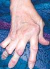
Advanced RA is characterized by severe cartilage and bone destruction (such as marginal joint erosions) that results in joint deformity (eg, subluxation, ulnar deviation, and bony ankylosis) (Figure 3). Inflammatory joint symptoms appear to be the main determinants of disability early in the disease, while joint destruction, measured radiographically, dominates late disease.
The cartilage-destroying mechanisms that produce joint destruction are activated very early in the course of the disease. Up to 93% of patients who have had RA for less than 2 years may manifest radiographic abnormalities.5,6 Some patients with a 4-month history of symptoms and normal radiographs have joint erosions detectable on MRI.7 Radiographic progression continues over a lifetime.
The systemic consequences of RA may also be severe. Multiple organ systems may be involved.1 Patients with RA have a 27% higher early mortality than the general population. They are twice as likely to suffer a myocardial infarction and are at significantly increased risk for a stroke or infection. They are also at risk for lymphoma. Negative effects on quality of life include pain associated with functional disability, chronic severe fatigue, depression, and loss of job productivity.
PATHOGENESIS
RA is an immune-mediated disease that occurs in a genetically predisposed host.1 It is characterized by the development of a hyperplastic synovial membrane (ie, pannus) that has been infiltrated by CD4+ T cells and macrophages, which eventuates in the production and release of proinflammatory cytokines. These events result in further localized and systemic inflammation; synoviocyte, chondrocyte, and osteoclast activation; and cartilage and bone destruction. In diseases such as RA, an imbalance develops between the proinflammatory cytokines (eg, interleukin [IL]-1b, tumor necrosis factor [TNF]-α) and the anti-inflammatory modulators (eg, soluble TNF receptors and IL-1 receptor antagonists). There are numerous other cytokines located in the RA synovial tissue or fluid; however, IL-1 and TNF appear to play a dominant role.
Because TNF triggers production of many other cytokines, including IL-1, it is generally viewed as pivotal in initiating the inflammatory response that causes cartilage and bone destruction and other systemic events.
MANAGEMENT
The current focus on the delivery of optimal patient-centered care has shifted emphasis away from management of the constitutional symptoms of RA to changing the course of the disease.
Current pharmacologic options include:
- Cyclooxygenase (COX)-2 inhibitors and NSAIDs. These agents control signs and symptoms, reduce acute inflammation, and provide moderate pain relief. They may serve as initial therapy for patients with nonspecific inflammation until a diagnosis is made and also as an adjunct to DMARDs for pain relief in severe cases.
- Corticosteroids. The anti-inflammatory and immunoregulatory activities of these agents help alleviate symptoms but have only nominal disease-modifying properties. Corticosteroids are more powerful anti-inflammatory agents than NSAIDs and COX-2 inhibitors, but long-term side effects of higher doses are problematic. Low-dose corticosteroids can be used in combination with DMARDs.
- DMARDs, such as methotrexate, sulfasalazine, leflunomide, and anti-TNF biologics, not only control signs and symptoms of RA but also retard disease progression, improve function, and enhance quality of life.8,9
Heightened awareness of the importance of early diagnosis, coupled with early intervention with DMARDs, offer hope of significant long-term benefit. To facilitate this process, a number of European and American rheumatologists, in conjunction with primary care physicians, have established interactive “early arthritis” clinics to facilitate prompt diagnosis and to overcome the obstacles to optimal care.10-12 These clinics are designed to provide urgent appointments for patients with suspected early RA.
The development of special educational programs is another strategy to improve collaboration between primary care clinicians and rheumatologists. The optimal treatment strategy is based on understanding the determinants of disease outcome. These include a family history of severe arthritis, early onset of severe synovitis with functional limitation, persistent elevation of ESR or C-reactive protein (CRP), or anti-CCP antibodies, and the presence of extra-articular manifestations. Other risk factors include poor socioeconomic status, low educational achievement, health-related beliefs that interfere with seeking medical attention, and inadequate access to health care.
A complete treatment program includes occupational and physical therapy and the use of educational resources and self-help programs sponsored by the Arthritis Foundation.The foundation’s Web site address is www.arthritis.org.
MAKING A DIAGNOSIS
There appears to be a window of opportunity for highly successful treatment of RA in the first year after diagnosis; the first 3 months of therapy are especially critical.10-12 There is also evidence that collaboration between primary care physicians and rheumatologists leading to an early referral can improve the long-term outcome of RA. Emery and colleagues10 suggest that rapid referral is warranted for patients with persistent pain and any of the following:
- 3 or more swollen joints.
- MTP/MCP joint involvement.
- Morning stiffness that lasts 30 minutes or more.
Cush11 points out that the accuracy of this approach can be enhanced by finding:
- Chronicity (duration of symptoms for more than 6 weeks; symptoms lasting more than 12 weeks are even more predictive of RA).
- Elevated acute phase reactants (ESR, CRP) or serologic abnormalities (RF, anti-CCP).

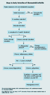
This information can be combined with an evidence-based algorithm that describes steps in the early detection of RA.13 Here I apply this approach to the case of the 38-year-old African American woman with joint pain whose case appears at the beginning of this article.
The initial assessment is directed toward determining whether symptoms are local or widespread and whether they suggest systemic illness. (For example, the patient in our case complained of fatigue.) Is the pain secondary to an articular or extra-articular event? Pain localized to the PIP, MCP, and MTP joints that is present during both active and passive motion is consistent with an articular disorder.
Close questioning of the patient about when the pain first started indicated that the onset had been approximately 7 weeks earlier. These findings indicate a chronic articular disorder (ie, lasting 6 or more weeks). However, many systemic diseases present with chronic polyarthritis; therefore, a complete history and physical examination are required.
It is also important to determine whether the patient’s chronic polyarthritis is inflammatory or noninflammatory. The initial history and physical findings that supported inflammation in our patient included morning stiffness, joint swelling, and pain. Laboratory findings that further supported inflammation included an elevated ESR. At this stage, therefore, the patient appeared to have a chronic inflammatory polyarthritis limited to the peripheral joints (Table 3).
The next step is to determine whether the polyarthritis is symmetric. Our patient's PIP, MCP, and MTP joints were affected bilaterally; this presentation is a characteristic symmetric pattern, commonly seen in RA, that spares the distal interphalangeal joints.
To establish a definitive diagnosis of RA, 4 of 7 criteria, as outlined by the ACR, are necessary (see Table 1). Before our patient saw the rheumatologist, only 3 of 7 classification criteria had been met (the presence of arthritis for 6 or more weeks involving 3 or more joints in a symmetric distribution). Findings on radiographs and results of tests for RF were initially negative, morning stiffness lasted only 30 minutes, and there were no rheumatoid nodules. Therefore, a presumptive diagnosis of early RA was made.
In addition to early RA, the differential diagnosis (based on age and sex) includes:
- Systemic lupus erythematosus (SLE). This multisystem disease occurs predominantly in women, particularly African Americans and Asians. It is characterized by arthritis with a joint distribution resembling that of RA. Our patient denied other signs and symptoms of SLE, including photosensitivity, rash, seizures, and dyspnea. Most important, the results of an antinuclear antibody assay-which has a sensitivity of 97% for SLE-were negative. SLE is, therefore, highly unlikely.
- Infection. Viral infection (particularly parvovirus infection) and subacute bacterial endocarditis have been associated with a chronic arthritis that resembles RA. Because the patient denied recent infection and did not demonstrate any symptoms that would suggest infection, an infectious cause of her joint pain is not likely. This conclusion was supported by a negative test for parvovirus antibodies.
- Undifferentiated seronegative polyarthritis. This condition is usually inflammatory, but it is not associated with increased titers of RF and is, therefore, atypical of RA.
After 4 months, the patient visited the rheumatologist. At that time, her symptoms were much worse. She now had morning stiffness lasting for 2 hours. RF and anti-CCP antibody test results were positive, hemoglobin levels had decreased, and the ESR was further elevated. Radiographic studies also revealed periarticular osteopenic changes in some MCP and PIP joints. Because these observations of clinical signs, symptoms, and laboratory results meet 6 of 7 criteria for establishing RA (only 4 of 7 are required), a definitive diagnosis was made. Note, however, that the ACR criteria for a definitive diagnosis of RA have a high sensitivity for moderate and advanced RA but are ineffective for diagnosing early RA.
More recently, evidence has been presented that, in conjunction with clinical symptoms, abnormal results of tests that measure both RF and anti-CCP antibodies may be helpful in identifying early RA (Table 4).14 In this study, in patients with synovitis of less than 3 months’ duration, the combined results of anti-CCP antibodies and RF have a high specificity and positive predictive value for the development of persistent RA.14 These observations, if confirmed in larger studies, suggest that this combination could help identify those patients most likely to benefit from very early DMARD intervention.
It is also important to note that other imaging techniques, such as contrast-enhanced MRI and color Doppler ultrasonography show promise for early detection of joint erosions and synovial inflammation.15
A SUMMARY OF THE EVIDENCE
Studies of RA provide evidence that:
- Damage to joints occurs very early and is progressive.
- Inflammatory symptoms do not correlate with joint destruction, but radiographic studies do
- DMARDs are effective in retarding joint destruction.
- Early DMARD intervention leads to better long-term outcomes.
- Early and aggressive therapy leads to the best outcomes.
- There is a “window of opportunity” for highly successful treatment of RA, especially in the first 3 months of therapy.
For these reasons, early diagnosis and DMARD therapy have become essential in achieving optimal patient outcomes. Because we primary care physicians are usually the first clinicians to see patients with persistent polyarthritis, it is imperative that we hone our skills in identifying clinical symptoms associated with early RA and that we partner with rheumatologists to promptly implement the most appropriate therapeutic intervention.
References:
REFERENCES:1. Goronzy JJ, Weyand CM. Rheumatoid arthritis: epidemiology, pathology, and pathogenesis. In: Klippel JH, Crofford LJ, Stone JH, Weyand CM, eds. Primer on the Rheumatic Diseases. 12th ed. Atlanta: Arthritis Foundation; 2001:209-225.
2. Gamez-Nava JI, Gonzalez-Lopez L, Davis P, Suarez-Almazor ME. Referral and diagnosis of common rheumatic diseases by primary care physicians. Br J Rheumatol. 1998;37:1215-1219.
3. Yelin EH, Such CL, Criswell LA, Epstein WV. Outcomes for persons with rheumatoid arthritis with a rheumatologist versus a non-rheumatologist as the main physician for this condition. Med Care. 1998;36:513-522.
4.Pincus T, Gibofsky A, Weinblatt ME. Urgent care and tight control of rheumatoid arthritis as in diabetes and hypertension: better treatments but a shortage of rheumatologists. Arthritis Rheum. 2002;46:851-854.
5. van der Heijde DM, van Leeuwen MA, van Riel PL, van de Putte LB. Radiographic progression on radiographs of hands and feet during the first 3 years of rheumatoid arthritis measured according to Sharp's method (van der Heijde modification). J Rheumatol. 1995;22:1792-1796.
6. Fuchs HA, Kaye JJ, Callahan LF, et al. Evidence of significant radiographic damage in rheumatoid arthritis within the first 2 years of disease. J Rheumatol. 1989;16:585-591.
7. McQueen FM, Stewart N, Crabbe J, et al. Mag-netic resonance imaging of the wrist in early rheu-matoid arthritis reveals a high prevalence of erosions at four months after symptom onset. Ann Rheum Dis. 1998;57:350-356.
8. Lard LR, Visser H, Speyer I, et al. Early versus delayed treatment in patients with recent-onset rheumatoid arthritis: comparison of two cohorts who received different treatment strategies. Am J Med. 2001;111:446-551.
9. Landewe RB, Boers M, Verhoeven AC, et al. COBRA combination therapy in patients with early rheumatoid arthritis: long-term structural benefits of a brief intervention. Arthritis Rheum. 2002;46: 347-356.
10. Emery P, Breedveld FC, Dougados M, et al. Early referral recommendation for newly diagnosed rheumatoid arthritis: evidence based development of a clinical guide. Ann Rheum Dis. 2002;61:290-297.
11. Cush J. Early arthritis clinics: if you build it will they come? J Rheumatol. 2005;32:203-207.
12. Nell VP, Machold KP, Eberl G, et al. Benefit of very early referral and very early therapy with disease-modifying anti-rheumatic drugs in patients with early rheumatoid arthritis. Rheumatology. 2004;43:906-914.
13. Ellrodt AG, Cho M, Cush JJ, et al. An evidence-based medicine approach to the diagnosis and management of musculoskeletal complaints. Am J Med. 1997;103:3S-6S.
14. Raza K, Breese M, Nightingale P, et al. Predictive value of antibodies to cyclic citrullinated peptide in patients with very early inflammatory arthritis. J Rheumatol. 2005;32:231-238.
15. Ostergaard M, Ejbjerg B. Magnetic resonance imaging of the synovium in rheumatoid arthritis. Semin Musculoskelet Radiol. 2004;8:287-299.
16. Diffuse connective tissue disease. In: The Merck Manual of Diagnosis and Therapy. 17th ed. Whitehouse Station, NJ: Merck Research Laboratories; 1999:417-423.
17. The University of Texas Southwestern Medical Center at Dallas. Algorithms for the diagnosis and management of musculoskeletal complaints. 1996. Available at: http://ww3.utsouthwestern.edu/cme/ endurmat/lipsky/index.html. Accessed April 7, 2005.
