- Clinical Technology
- Adult Immunization
- Hepatology
- Pediatric Immunization
- Screening
- Psychiatry
- Allergy
- Women's Health
- Cardiology
- Pediatrics
- Dermatology
- Endocrinology
- Pain Management
- Gastroenterology
- Infectious Disease
- Obesity Medicine
- Rheumatology
- Nephrology
- Neurology
- Pulmonology
Pneumatosis Intestinalis Triggers Pneumoperitoneum
Pneumatosis cystoides intestinalisa rare condition, is characterized by gaseous cysts within the submucosal and subserosal spaces of the bowel wal

Figure 1
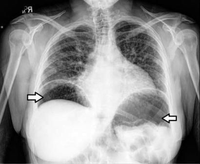
A 53-year-old white woman with schizophrenia was transferred from a psychiatric center after vague complaints of shortness of breath led to obtaining a chest x-ray film, which revealed significant subdiaphragmatic free air (Figure 1). On admission, the patient was alert and oriented; she insisted she was medically healthy and admitted only to mild abdominal distention. She denied current shortness of breath or any other GI complaints and further denied recent fever or chill. Her past history was significant for scleroderma and uncontrolled schizophrenia that resulted from nonadherence to prescribed aripiprazole.
On initial examination, the patient was alert and cooperative, appeared well with stable vitals, and had no abnormal cardiopulmonary findings. Her abdominal examination showed diffuse distention and tympany, minimal tenderness to palpation, normal bowel sounds, and no guarding or rebound. Her only other significant physical findings were sclerodactyly and diffuse xerosis. The findings from her complete blood cell count, complete metabolic panel, and urinalysis were within normal limits; her toxicology screen result was negative.
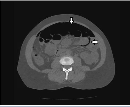
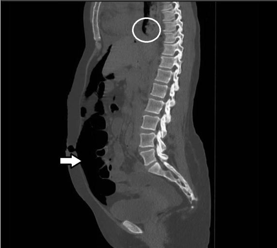
CT scans of the patient’s chest, abdomen, and pelvis further detailed the pneumoperitoneum and showed intraluminal free air alongside gravity-defying intramural air in the small intestine, although there were no signs of perforation (Figure 2). Of note, the radiographs revealed esophageal stricture with proximal dilatation in addition to pneumoperitoneum, consistent with the patient’s diagnosis of scleroderma (Figure 3).
Diagnosis
Radiographic findings of air collections within the wall of the patient’s intestines established pneumatosis cystoides intestinalis (PCI) as the cause of her spontaneous pneumoperitoneum. The combination of a benign abdomen on examination, the absence of any other explanation for pneumoperitoneum, and the presence of the typical findings for PCI in a patient with scleroderma fully supported this diagnosis.

Figure 2

Figure 3
Discussion
PCI, a rare condition, is characterized by gaseous cysts within the submucosal and subserosal spaces of the bowel wall.1 It is less a disease than a radiologic or endoscopic finding. PCI can be idiopathic or, more frequently, the result of numerous medical conditions that theoretically increase the possibility of introducing air into the abdominal cavity (Table).2,3
On the basis of cadaver studies, the overall incidence of PCI in the general population is estimated to be 0.3%. PCI is seen more frequently in men, with a peak incidence between ages 30 and 50 years.4
Patients with PCI may present asymptomatically after radiographic or endoscopic discovery. Or, they may exhibit a variety of GI symptoms, including nausea, vomiting, diarrhea, constipation, abdominal pain and, less frequently, weight loss, bloody stools, and tenesmus.
The diagnosis of PCI is primarily radiological; the test of choice is abdominal CT scan. Common findings include linear or bubbly intramural gas patterns parallel to the bowel wall, often gravity-defying and in circular arrangement on cross-section, as was seen on our patient’s CT scan. Positive radiological findings are lacking in about one-third of patients; follow-up histology can confirm the diagnosis but usually is unnecessary.4
Treatment
Treatment for patients with PCI varies with different patient presentations. When a patient presents with PCI as an incidental finding, treatment may be controversial, often resulting in expectant management. When a patient presents with intra-abdominal free air or GI symptoms, PCI typically is managed conservatively with bowel rest, intravenous fluids and, optionally, corticosteroids, laxatives, elemental diets, broad-spectrum antibiotics, and hyperbaric oxygen.5
Inspiration of high-flow oxygen by face mask has been shown to help eliminate the cysts, but discontinuing therapy may lead to recurrence.3 Surgery is required only for the infrequent but urgent complications of PCI, such as mesenteric ischemia, perforation, peritonitis, and abdominal sepsis.4
In our patient’s case, surgeons were consulted initially in expectation of perforated bowel, but they concurred with the nonoperative diagnosis. Conservative management was initiated that consisted of bowel rest, intravenous fluids, and serial monitoring of vital signs.
After a brief period of uneventful observation, the patient’s diet was restarted and she made a complete recovery. She was then transferred back to the psychiatric center for continued management of schizophrenia and outpatient management of her scleroderma.
Summary
PCI remains an often-overlooked cause of spontaneous pneumoperitoneum. Although the majority (more than 90%) of cases of pneumoperitoneum arise from an intra-abdominal perforation and require surgical repair, PCI is one of the few nonsurgical etiologies that may be managed with conservative therapies as outlined above.2
Indeed, because the iatrogenic risks of unnecessary surgery are so consequential, clinicians must investigate PCI as a potential cause of intraperitoneal free air when the patient’s initial clinical picture is suggestive of a benign process. We present a case of PCI causing pneumoperitoneum to raise clinician awareness of this disorder and thereby help prevent unnecessary surgical intervention.
References:
1. Vischio J, Matlyuk-Urman Z, Lakshiminarayanan S. Benign spontaneous pneumoperitoneum in systemic sclerosis. J Clin Rheumatol. 2010;16:379-381.
2. Williams NM, Watkin DF. Spontaneous pneumoperitoneum and other nonsurgical causes of intraperitoneal free gas. Postgrad Med J. 1997;73:531-537.
3. Vogel Y, Buchner NJ, Szpakowski M, et al. Pneumatosis cystoides intestinalis of the ascending colon related to acarbose treatment: a case report. J Med Case Rep. 2009;3:9216.
4. Bartnik W, Pachlewski J, Regula J. Colorectal disorders. In: Classen M, Tytgat G, Lightdale C, eds. Gastroenterological Endoscopy. 2nd ed. New York: Thieme Medical Publishers; 2010:596-615.
5. Arikanoglu Z, Aygen E, Camci C, et al. Pneumatosis cystoides intestinalis: a single center experience. World J Gastroenterol. 2012;18:453-457.
