- Clinical Technology
- Adult Immunization
- Hepatology
- Pediatric Immunization
- Screening
- Psychiatry
- Allergy
- Women's Health
- Cardiology
- Pediatrics
- Dermatology
- Endocrinology
- Pain Management
- Gastroenterology
- Infectious Disease
- Obesity Medicine
- Rheumatology
- Nephrology
- Neurology
- Pulmonology
Painful Oral Lesions: What to Look For, How to Treat, Part 2
ABSTRACT: Painful recurrent ulceration of gingival tissue suggests a secondary intraoral presentation of herpes simplex virus (HSV) infection. Unlike the lesions of HSV, lesions associated with coxsackievirus do not erupt in the anterior mouth but rather on the soft palate and pharynx. Furthermore, unlike HSV infection, coxsackie infections may recur, because there is considerable viral variation. Patients with atrophic or erythematous candidiasis report burning pain and a metallic taste. The typical patient with benign mucous membrane pemphigoid is a woman older than 50 years; the condition usually involves the attached gingiva around the teeth. The lesions of erythema multiforme may erupt on any intraoral mucosa; biopsy may be required to rule out other conditions with similar presentations.
In this second article in my 2-part series on painful oral lesions, I focus on how to identify and treat nonmalignant disorders, including viral infections, candidiasis, benign mucous membrane pemphigoid (BMMP), and erythema multiforme. Although the diagnosis can usually be made based on oral examination findings coupled with the patient's history, I will also discuss the settings in which further workup is required. In a previous article (CONSULTANT, November 2006, page 1497), I reviewed the diagnosis and treatment of oral cancer and lichen planus.

Figure 1

Figure 2
VIRAL INFECTIONS
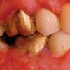
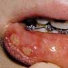
Herpes simplex. The 2 most common infectious conditions in the general practice setting are herpes simplex virus (HSV) infection (type 1 and, less frequently, type 2) (Figure 1) and fungal disease. These conditions are not generally mistaken for each other because their clinical presentations are quite distinct.HSV infection is sometimes confused with aphthous stomatitis (a noninfectious, immunologic-mediated condition precipitated by stress, food sensitivities, trauma, and endocrine disorders) because both of these conditions are characterized by multiple oral ulcerations. However, patients with primary HSV infection--unlike those with aphthous stomatitis (Figure 2)--present with extraoral symptoms including irritability, malaise, headache, and low-grade fever. Further, multiple pinhead lesions are observed on the attached gingiva surrounding the teeth and on the palate, 2 areas not affected in aphthous stomatitis. Both conditions are painful, but pain is generally more severe in patients with herpes simplex. Patients may not be adequately hydrated because drinking is painful. Examination reveals distinct halitosis and submandibular lymphadenitis.1 Both HSV infection and aphthous stomatitis are self-limited, but HSV infection has been associated with morbidity and, in rare cases, mortality.2

Figure 3
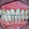
In adults, painful ulceration of gingival tissue that recurs in the same location suggests a secondary intraoral presentation of HSV infection--recurrent intraoral herpes (Figure 3) (the intraoral equivalent of the lip lesion). Although recurrent HSV lesions usually arise in the same location, they may occur on nongingival mucosa, especially in immunocompromised persons.3 HSV-2 also recurs intraorally,4 but not as frequently as HSV-1. A simple assessment of suspected recurrent intraoral HSV infection may be done through viral culture. Cytologic evaluation with fluorescent staining is rarely used but it may be helpful if available; antibody titers can provide additional evidence of disease. In immunocompromised persons, it may be prudent to perform cultures of any intraoral lesions for HSV, regardless of location, because early intervention helps prevent morbidity.5
Therapy for primary or recurrent intraoral HSV infection includes topical and systemic anesthetics and analgesics; supportive therapy, including rest and fluid and food consumption; and vitamin and mineral supplementation. Oral acyclovir can reduce symptom duration but is recommended only in cases of underlying immunosuppression because of the risk of resistance.6 However, in immunosuppressed patients who are undergoing treatment (including bone marrow transplantation) for myeloid leukemia, the use of acyclovir or another antiviral is recommended because these agents significantly reduce or prevent recurrent oropharyngeal HSV infection, reduce the duration of leukopenia, and reduce the number of days with fever.7 Valacyclovir, which has greater bioavailability than acyclovir, suppresses recurrent herpes labialis in immunocompromised patients.8 It has also been recommended to prevent recrudescence in immunocompetent persons with a history of HSV disease who require dental treatment.9
Other herpesvirus infections. Other human herpesviruses that can cause intraoral ulceration include:
- Epstein-Barr, implicated in the development of hairy leukoplakia seen in HIV-infected patients.
- Cytomegalovirus, found primarily in immunocompromised persons and in some patients with salivary disease.
- HSV-8,10 associated with Kaposi sarcoma and HIV infection.
- Varicella-zoster virus.
Coxsackievirus infections. The coxsackieviruses cause several mildly painful intraoral ulcerative conditions, including herpangina, hand-foot-mouth disease, and lymphonodular pharyngitis. Unlike the lesions of HSV, lesions associated with coxsackievirus do not occur in the anterior mouth but rather on the soft palate and pharynx. Furthermore, unlike HSV infection, coxsackie infections may recur, because there is considerable viral variation. Coxsackie-induced disease, typically seen in children, is self-limited and may be treated in the same manner as primary HSV infection.
CANDIDIASIS
This is the most common intraoral fungal infection. Candidiasis may also cause or contribute to fissuring at the corner of the mouth (cheilitis or cheilosis).
Pseudomembranous candidiasis is easy to recognize because of the large white plaques that can be easily removed by a swab (leaving a painful bleeding subsurface), but the chronic, atrophic, hyperplastic, and erythematous forms are more difficult to differentiate from other conditions, such as erosive lichen planus. Patients with atrophic or erythematous candidiasis may describe burning pain and a metallic taste. Definitive diagnosis of these disease variations and differentiation from noncandidal infection (as well as from other fungal infections) may require biopsy. Consider candidiasis when a presumed staphylococcal or streptococcal oral infection fails to improve with antibiotics. A response to antifungal therapy strongly supports the diagnosis.
Options for the treatment of intraoral candidiasis include topical antifungal agents such as nystatin suspensions or pastilles or clotrimazole troches (10 mg). Effective systemic medications include ketoconazole (200 mg/d for 14 days) and fluconazole (100 mg bid for 14 days). Recurring candidiasis despite adequate pharmacologic management may indicate immunosuppression or myelosuppression and requires additional evaluation. In patients with a denture or partial denture, it may be helpful to disinfect the prosthesis with denture-soaking solution and apply an antifungal powder or cream to the tissue-contacting surface before insertion.

Figure 4
BMMP
This rare condition, the result of autoimmune humoral abnormality that alters basal cell adhesion, causes gingival vesicles that subsequently slough, leaving localized erosion, erythema, and pain (Figure 4). The typical patient is a woman older than 50 years. Although BMMP usually involves the attached gingiva around the teeth, lesions may also occur on the cheeks and palate. Because vesicle formation is subepithelial, it is common to find unruptured vesicles, a feature that helps to differentiate this condition from pemphigus.11 Oral examination usually reveals a positive Nikolsky sign (the sloughing of tissue when it is touched lightly).
Lesions may affect other squamous epithelia, including those of the conjunctiva, vulva, penis, and larynx. Most worrisome is the possibility of conjunctival blistering followed by scarring in patients with oral BMMP, because this can cause significant vision impairment or blindness. The diagnosis of BMMP is aided by biopsy and immunofluorescence study, as this technique easily differentiates the condition from lichen planus and pemphigus.12
The differential diagnosis of BMMP includes pemphigus vulgaris, bullous pemphigoid, erythema multiforme, and erosive lichen planus. Management of BMMP typically involves topical and systemic anti-inflammatory and immunosuppressive agents. Dapsone may be effective in patients who cannot tolerate these agents or when a reduced dosage is necessary.13

Figure 5
ERYTHEMA MULTIFORME
Erythema multiforme (EM) (Figure 5) is an inflammatory disorder caused by immune complexes that filter out in vessel walls of the submucosa and initiate neutrophil and macrophage migration.14 The result is tissue destruction in the form of bullae, mild or severe epithelial erosion, and painful ulceration. Lesions may erupt on any intraoral mucosa, particularly on the cheek region, soft palate, vestibule, and lips. The ulcers are irregular in shape, shallow, and covered by a gray/yellow pseudomembrane.
Three variations of EM have been described:
- EM minor is usually secondary to HSV-1 infection.
- EM major is a reaction to a number of drugs, chiefly sulfur-based compounds (sulfa antibiotics and hypoglycemic sulfonylureas).
- Stevens-Johnson syndrome is associated with the sulfur-based compounds as well as the penicillins.15,16 An association with sulfur-based food additives has not been conclusively demonstrated.
The lesions of EM are quite distinctive. There may be erythematous patches or a bull's-eye pattern, with concentric red rings surrounding a central circular erythematous area involving the keratinized skin. Clinically, a drug or HSV history is the most important factor in establishing the diagnosis and differentiating EM from other vesiculobullous diseases. Because clinical findings are nonspecific, biopsy may be required to help rule out other mucocutaneous conditions with a similar presentation, such as paraneoplastic pemphigus, erosive lichen planus, pemphigus, and BMMP.
Treatment of EM not associated with concurrent HSV infection depends on the severity of the disease. In some cases, a 6-day course of high-dose corticosteroids may be sufficient. For resistant disease, a 2- to 4-week course of high-dose corticosteroids with taper may be required. An antiviral agent can be added for associated HSV or if there is concern about recrudescence. In one report, dapsone was used successfully in a patient with refractory disease.17
Secondary infection may require treatment with a topical antibiotic, such as tetracycline, coupled with low- to moderate-dose analgesics. Stevens-Johnson syndromeproduces severe ocular, nasal, pharyngeal, laryngeal, anogenital and upper respiratory lesions, as well as dermal abnormality, and may require inpatient management.
References:
REFERENCES: 1. Kolokotronis A, Doumas S. Herpes simplex virus infection, with particular reference to the progression and complications of primary herpetic gingivostomatitis. Clin Microbiol Infect. 2006;12:202-211.
2. Tyler K. Herpes simplex virus infections of the central nervous system: encephalitis and meningitis, including Mollaret's. Herpes. 2004;11(suppl 2): 57A-64A.
3. Eisen D. The clinical characteristics of intraoral herpes simplex virus infection in 52 immunocompetent patients. Oral Surg Oral Med Oral Pathol Oral Radiol Endod. 1998;86:432-437.
4. Glick M. Clinical aspects of recurrent oral herpes simplex virus infection. Compend Contin Educ Dent. 2002; 23(7, suppl 2):2-4.
5. Woo SB, Lee SF. Oral recrudescent herpes simplex virus infection. Oral Surg Oral Med Oral Pathol Oral Radiol Endod. 1997;83:239-243.
6. Shillitoe E. Herpes simplex virus. In: Millard HD, Mason DK, eds. The Second World Workshop on Oral Medicine. Ann Arbor, Mich: University of Michigan Press; 1993.
7. Hann IM, Prentice HG, Blacklock HA, et al. Acyclovir prophylaxis against herpes virus infection in severely immunocompromised patients: randomized double-blind trial. Br Med J (Clin Res Ed). 1983;287: 384-388.
8. Baker D, Eisen D. Valacyclovir for prevention of recurrent herpes labialis: 2 double-blind, placebo-controlled studies. Cutis. 2003;71:239-242.
9. Miller CS, Cunningham LL, Lindroth JE, Avdiushka SA. The efficacy of valacyclovir in preventing recurrent herpes simplex virus infections associated with dental procedures. J Am Dent Assoc. 2004;135:1311-1318.
10. Schulz TF. The pleiotropic effects of Kaposi's sarcoma herpesvirus. J Pathol. 2006;208:187-198.
11. Scully C, Carrozzo M, Gandolfo S, et al. Update on mucous membrane pemphigoid: a heterogeneous immune-mediated subepithelial blistering entity. Oral Surg Oral Med Oral Pathol Oral Radiol Endod. 1999;88:56-68.
12. Carrozzo M, Broccoletti R, Carbone M, et al. Pemphigoid of the mucous membranes. The clinical, histopathological and immunological aspects and current therapeutic concepts. Minerva Stomatol. 1996;45:455-463.
13. Ciarrocca K, Greenberg M. A retrospective study of the management of oral mucous membrane pemphigoid with dapsone. Oral Sur Oral Med Oral Pathol Oral Radiol Endod. 1999;88:159-163.
14. Greenberger PA. Drug allergy. J Allergy Clin Immunol. 2006;117(2 suppl Mini-Primer): S464-S470.
15. Ayangco L, Rogers RS 3rd. Oral manifestations of erythema multiforme. Dermatol Clin. 2003;21: 195-202.
16. Forman R, Koren G, Shear NH. Erythema multiforme, Stevens-Johnson syndrome and toxic epidermal necrolysis in children: a review of 10 years' experience. Drug Saf. 2002;25:965-972.
17. Hoffman LD, Hoffman MD. Dapsone in the treatment of persistent erythema multiforme.J Drugs Dermatol. 2006;5:375-376.
AI-Powered Diabetes Prevention Program Intervention Matches Human Coaching in Landmark Trial
October 27th 2025After 1 year of coaching by the AI app or a DPP-associated coach, reductions in weight and HbA1c and increases in physical activity were equal, an outcome with great promise for scalability.
