- Clinical Technology
- Adult Immunization
- Hepatology
- Pediatric Immunization
- Screening
- Psychiatry
- Allergy
- Women's Health
- Cardiology
- Pediatrics
- Dermatology
- Endocrinology
- Pain Management
- Gastroenterology
- Infectious Disease
- Obesity Medicine
- Rheumatology
- Nephrology
- Neurology
- Pulmonology
Overuse Injury: Is It Tendonitis or Bursitis?
A 36-year-old man presented with a 2-week history of pain, swelling, and redness of the right elbow. He also had decreased range of motion during elbow flexion. There was no associated fever or recent illness.
A 36-year-old man presented with a 2-week history of pain, swelling, and redness of the right elbow. He also had decreased range of motion during elbow flexion. There was no associated fever or recent illness. Before the symptoms developed, he had fallen while playing basketball and hit his elbow on the court.
On examination, active range of motion was reduced in all directions. However, there was more limitation in flexion because of pain. The pain was localized to the posterior aspect of the elbow where the swelling and erythema were most prominent. The olecranon area was tender to palpation. Standard radiographs of the elbow were normal.
Do you suspect tendonitis or bursitis in this patient?
Tendonitis and bursitis can have similar presentations, and both can result from repetitive movements. Here we offer tips on distinguishing tendonitis from bursitis (Table 1). We describe clues in the history, key physical findings, and tests that can help clinch the diagnosis. We also discuss effective treatment approaches.
TENDONITIS
Because most symptomatic disorders that affect tendons are not inflammatory, some experts suggest that the generic term "tendinopathy" may be more accurate than "tendonitis."1 The origin of a tendinopathy is likely multifactorial and may include overuse, inflexibility, equipment problems, age-related tendon degeneration, and biomechanical functions.2 Commonly affected areas include the following:

• Rotator cuff of the shoulder (supraspinatus tendinopathy).
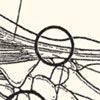
• Elbow (lateral epicondylitis, or "tennis elbow," and medial epicondylitis, or "golfer's elbow") (Figure 1).
• Knee (eg, patellar tendonitis and iliotibial band syndrome).
• Lower leg (anterior tibial tendonitis [ie, "shin splints"]).
• Heel (Achilles tendonitis).
Clinical features. Typically, patients with a tendinopathy have started a new activity or increased the intensity of a current activity before the onset of symptoms. Initially, pain may occur with activity and then diminish after a warm-up period. The pain progressively increases in intensity and duration; in later stages, it may occur at rest. The pain is described as "sharp" or "stabbing" during the aggravating activity and as a "dull ache" immediately after activity or at rest.
Physical examination. When a patient presents with symptoms that suggest a tendinopathy, inspect the affected area for muscle atrophy, asymmetry, swelling, erythema, and joint effusions. Muscle atrophy can be an important clue to the duration of the tendinopathy, because it is often associated with chronic disorders. Swelling, erythema, and asymmetry are common physical findings. Joint effusions are uncommon with tendinopathy and suggest a possible intra-articular pathology.1
Range of motion is often restricted on the pathological side. Palpation elicits pain comparable to that felt during activity.

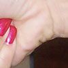
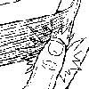
Clinical diagnostic tests. The clinical diagnosis of tendinopathy is supported by physical maneuvers that promote tendon loading and reproduce the patient's pain. Such maneuvers involve passively stretching an affected tendon (eg, the Finkelstein test, which can reproduce the pain of de Quervain tenosynovitis, Figure 2) or asking the patient to actively push against resistance (eg, the Jobe test, which can reproduce the pain of supraspinatus tendinopathy). In the Jobe test, both arms are abducted to 90 degrees and held fully pronated slightly in front of the body. The patient is asked to hold his or her arms in this position while the clinician attempts to force the arms downward. The test is positive when the patient is unable to hold up the arm or has decreased strength with pain. Figure 3 shows how the pain of lateral epicondylitis can be reproduced during resisted extension of the patient's wrist.3 This pain can also be elicited when the patient exerts a forceful grip. Imaging studies. Plain radiographs usually do not show the soft tissue changes in tendinopathy. They may reveal bony abnormalities, such as loose bodies and osteoarthritis, that can cause similar symptoms. Ultrasonography and MRI can be useful in the evaluation of tendinopathies. In patients with chronic Achilles tendinopathy, for instance, ultrasonography can help ensure clinical diagnosis and guide corticosteroid injection.4 The effectiveness of treatment can be monitored with ultrasonography and pressure algometry or with MRI.5

BURSITIS
The bursal sac can become inflamed as a result of injury, overuse, infectious disease, or inflammatory disorders (such as arthritis).6 The following bursae most often become inflamed:
• Subdeltoid and subacromial.
• Olecranon.
• Ischial and trochanteric.
• Prepatellar.
Clinical characteristics. Patients with bursitis typically have swelling, erythema, and localized pain over the affected bursa when in motion and at rest. Range of motion may also be decreased. Intermittent loss of active movement but preserved passive movement is a characteristic finding; however, this is also common in patients with a tendinopathy.
Physical examination. When you suspect bursitis, ask patients about acute trauma, repetitive injury, or occupational or extracurricular activities that involve the affected area. Swelling, if present, is easier to detect in superficial bursae than in deeper bursae. Erythema could be a sign of cellulitis associated with an infected bursa, or septic bursitis. Symptoms suggestive of a current or recent bacterial infection or the presence of a fever strongly indicate septic bursitis.
Diagnosis. The diagnosis of bursitis is clinical. Further evaluation for infection or crystal-associated disease, such as gout or pseudogout, may be necessary based on the initial history and physical examination findings, especially if signs of inflammation are accompanied by effusion. The superficial bursae, including the olecranon, prepatellar, and infrapatellar bursae, are the most susceptible to infection because of the predisposition to trauma in these areas.7
Plain radiographs are usually normal in patients with bursitis. However, they can help rule out a fracture, dislocation, or other suspected pathology. Radiographs may show calcification of the bursa in patients with chronic bursitis or calcific bursitis. Soft tissue calcification may be caused by deposition of crystals of hydroxyapatite, basic calcium phosphate, or calcium pyrophosphate dihydrate.8
Ultrasonography may be required to guide the needle into the bursa (especially the deep bursae) during diagnostic aspiration or injection therapy. If necessary, MRI can help depict bursal/prebursal fluid, associated abscesses, and adjacent soft tissue structures.9
Aspiration of the bursa and fluid analysis can rule out septic bursitis and can be therapeutic. A complete blood cell count with differential, Gram stain, culture, and crystal analysis of the bursal fluid should be obtained. The white blood cell count in septic bursitis is lower than that of septic arthritis; a count between 5000/mL and 20,000/mL may indicate septic bursitis. Gram stain sensitivity in detecting organisms varies from 15% to 100%.7 Culture of the aspirate with liquid medium remains the definitive diagnostic test for septic bursitis.10
The patient in the opening case had olecranon bursitis. The diagnosis was based on the location of the pain, swelling, and erythema, which were over the bursa posterior to the olecranon process. The cause was likely related to his fall on the basketball court. Aspiration and analysis of bursal fluid showed no infection.
CONSERVATIVE TREATMENT
In most types of tendinopathy and bursitis, the first approach to treatment is conservative.
Tendinopathy. Relative rest of the affected area, ice, NSAIDs, and stretching and strengthening exercises are all suitable for patients with acute tendinopathy.
Rest. Relative rest of the affected area prevents repetitive loading that occurs with activity. It also prevents ongoing damage, reduces pain, and promotes healing. Recommendations for the duration of rest are unclear, and different regimens have not been studied in randomized controlled trials.1 Most clinicians advise patients to rest the affected area but continue activities that do not cause pain and avoid complete immobilization to prevent muscular atrophy and deconditioning.
Ice. During the first 24 to 48 hours of an injury, icing the affected area can help reduce swelling and pain associated with acute tendinopathy. One study concluded that the application of ice through a wet towel for 10-minute periods is most effective for acute soft tissue injuries.11NSAIDs. These medications can relieve tendinopathy pain. A typical regimen may include ibuprofen, 800 mg tid for 2 weeks, if there are no contraindications to high-dose NSAIDs. However, because the vast majority of tendinopathies are not inflammatory, whether NSAIDs are more effective than other analgesics is unclear.12 In a study of adults with lateral elbow pain, researchers were unable to recommend or discourage the use of oral NSAIDs in the short-term treatment of tendinopathies based on insufficient evidence.13
Strengthening and stretching exercises. Eccentric strength training is effective in treating tendinopathies and helps promote the formation of new collagen.14 Eccentric exercise has proved beneficial in Achilles tendinopathy and patellar tendinopathy and may be useful in other tendinopathies.15,16 Eccentric contraction involves lengthening of muscle fibers as the muscle contracts, preferentially loading the tendon.
Patients should begin strengthening and stretching exercises after the pain has abated. One eccentric exercise for Achilles tendinopathy is the toe raise, which is performed while standing on a step. The patient balances on the toes, then lifts the uninvolved foot and slowly lowers the other foot into dorsiflexion.
Orthotics and braces. Tennis elbow bands and shoe inserts are used to support, unload, and protect tendons during activity and can provide additional benefit when used with other therapies. Although few studies have evaluated their efficacy, orthotics and braces are safe, widely used, and often helpful in correcting associated biomechanical problems, such as excessive foot pronation and pes planus.15Bursitis. Conservative treatment of aseptic bursitis is similar to that of acute tendinopathy and includes relative rest, cold and heat treatments, elevation of the affected extremity, NSAIDs, and joint protection.
Ice therapy and NSAIDs are used initially to relieve acute symptoms of inflammation. Ibuprofen, 800 mg tid for 2 weeks, may be used, if there are no contraindications to high-dose NSAIDs.
Educate patients with bursitis on how to avoid aggravating their injury by not placing direct pressure on the affected area (eg, for elbow pain, avoid pushing up with the elbows; for knee pain, avoid kneeling; and for hip pain, avoid lying on the affected side). Provide patients with the appropriate method of joint protection for the affected area. For instance, patients at risk for prepatellar bursitis can use kneeling pads, and patients with Achilles bursitis can insert a thick heel pad in their shoe to raise the heel.
In the patient with olecranon bursitis, the region was immobilized for 7 days and activities that aggravated the symptoms were discontinued for 1 to 2 weeks. The patient was instructed on how to use ice compresses to help reduce the swelling. Ibuprofen (800 mg tid) with food for 2 weeks was prescribed for the pain. He was symptom-free after 3 weeks.
INJECTION THERAPY
Corticosteroid injections are generally reserved for patients who do not respond to conservative treatment. Local corticosteroid injections of the tendon is a topic of debate; currently, no evidence-based guidelines support their use in tendinopathy.1 Because corticosteroids may have harmful effects on the tendon, direct injection into the tendon should be avoided. The effects of peritendinous corticosteroid injections are unknown; however, they can be used with caution. Avoid repetitive corticosteroid injections in any site, because corticosteroids may inhibit healing and reduce the tensile strength of the tissue, which could lead to tendon rupture.1,17
In overuse injuries, corticosteroid injections should not replace discontinuation or modification of the offending activity. Corticosteroid injections should not be given if septic bursitis is suspected. Other contraindications to injection include local cellulitis, bacteremia, Achilles tendinopathies, and minimal response after 2 treatments.
A wide range of corticosteroids have been used. No single agent is demonstrably superior.1 Corticosteroids can be mixed in the same syringe with lidocaine and/or bupivacaine. Triamcinolone (Kenalog, 40 mg) mixed with an equal amount of 1% lidocaine has worked well in our practice. The recommended volume and doses for corticosteroid injection are listed in Table 2. Complications include a "steroid flare" (worsening of symptoms for a few days after injection), tenderness and erythema at the injection site, soft tissue atrophy, local infection, and tendon rupture.
References:
REFERENCES:
1.
Wilson JJ, Best TM. Common overuse tendon problems: a review and recommendations for treatment. Am Fam Physician. 2005;72:811-818.
2.
Almekinders LC, Weinhold PS, Maffulli N. Compression etiology in tendinopathy. Clin Sports Med. 2003;22:703-710.
3.
Rettig A. Tests and treatments of hand, wrist, and elbow overuse syndromes: 20 clinical pearls. J Musculoskel Med. 2003;20:136-146.
4.
Fredberg U, Bolvig L, Pfeiffer-Jensen M, et al. Ultrasonography as a tool for diagnosis, guidance of local steroid injection and, together with pressure algometry, monitoring of the treatment of athletes with chronic jumper's knee and Achilles tendinitis: a randomized double-blind, placebo-controlled study. Scand J Rheumatol. 2004;33:94-101
5.
Shalabi A. Magnetic resonance imaging in chronic Achilles tendinopathy. Acta Radiol Suppl. 2004;(432):1-45.
6.
Liang KP, Matteson EL. Bursitis: common condition, uncommon challenge. J Musculoskel Med. 2006;23:513-522.
7.
Small LN, Ross JJ. Suppurative tenosynovitis and septic bursitis. Infect Dis Clin North Am. 2005;19:991-1005.
8.
Molloy ES, McCarthy GM. Calcium crystal deposition diseases: update on pathogenesis and manifestations. Rheum Dis Clin North Am. 2006;32:383-400, vii.
9.
Beltran J. MR imaging of soft-tissue infection. Magn Reson Imaging Clin N Am. 1995;3:743.
10.
Stell IM, Gransden WR. Simple tests for septic bursitis: comparative study. BMJ. 1998;316:1877.
11.
Bleakley C, McDonough S, MacAuley D. The use of ice in the treatment of acute soft-tissue injury: a systematic review of randomized controlled trials. Am J Sports Med. 1998;32:251-261.
12.
Almekinders LC, Temple JD. Etiology, diagnosis, and treatment of tendonitis: an analysis of the literature. Med Sci Sports Exerc. 1998;30:1183-1190.
13.
Green S, Buchbinder R, Barnsley L, et al. Non-steroidal anti-inflammatory drugs (NSAIDS) for treating lateral elbow pain in adults. Cochrane Database Syst Rev. 2001;(4):CD003686.
14.
Ohberg L, Lorentzon R, Alfredson H. Eccentric training in patients with chronic Achilles tendinosis: normalized tendon structure and decreased thickness at follow up. Br J Sports Med. 2004;38:8-11.
15.
Alfredson H, Pietila T, Jonsson P, Lorentzon R. Heavy-load eccentric calf muscle training for the treatment of chronic Achilles tendinosis. Am J Sports Med. 1998;26:360-366.
16.
Cannell LJ, Taunton JE, Clement DB, et al. A randomized clinical trial of the efficacy of drop squats or leg extension/leg curl exercises to treat clinically diagnosed jumper's knee in athletes: pilot study. Br J Sports Med. 2001;35:60-64.
17.
Kapetanos G. The effect of the local corticosteroids on the healing and biomechanical properties of the partially injured tendon. Clin Orthop Relat Res. 1982;163:170-179.
18.
Shahady E. Woman with chronic wrist pain. Consultant. 2007;47:398-404.
19.
Sanchez-Yamamoto D, Bush TM. Bursitis and tendinitis: injection therapy basics. J Musculoskel Med. 2002;19:13-23.
