- Clinical Technology
- Adult Immunization
- Hepatology
- Pediatric Immunization
- Screening
- Psychiatry
- Allergy
- Women's Health
- Cardiology
- Pediatrics
- Dermatology
- Endocrinology
- Pain Management
- Gastroenterology
- Infectious Disease
- Obesity Medicine
- Rheumatology
- Nephrology
- Neurology
- Pulmonology
Osteomyelitis in a 75-Year-Old Heroin User
Presenting complaints were fever, nausea, and lower abdominal pain that worsened with walking. Osteomyelitis is not commonly included in a differential for abdominal pain. This case is different.
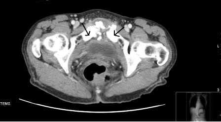
Figure. Click to enlarge.

A 75-year-old woman was admitted with complaints of fever, nausea, and lower abdominal pain that worsened with walking of a month’s duration. She denied any vomiting, constipation or diarrhea, gastrointestinal bleeding, chest pain, shortness of breath, palpitations, burning micturition, increased urinary frequency, hematuria or vaginal discharge. Her past medical history included hypertension, hyperlipidemia, and chronic untreated hepatitis B and C. She was an active intravenous heroin abuser. She denied smoking or drinking alcohol. Her medications included amlodipine, simvastatin, and aspirin. Her initial blood pressure was 140/74 mm Hg; heart rate, 116 beats/min regular; respiratory rate, 20 breaths/min; O2 saturation, 98% on ambient air; and temperature 101.2°F.
Physical examination was notable for pallor, diffuse tenderness over the lower abdomen, no guarding or rigidity, and normal bowel sounds. The balance of the physical examination, including rectal exam, was normal.
Relevant laboratory data included: hemoglobin, 11.4 g/dL (12-16 g/dL); hematocrit, 35.4% (38%-47%); WBC count, 18.62 (4.3-11 K/µL) with left shift; ESR, 102 mm/h (0-30 mm/h); CRP, 182.66 mg/L (<3 mg/L); normal renal and hepatic function; and a normal urinalysis. Blood cultures were positive for methicillin-resistant Staphylococcus aureus (MRSA).
Chest x-ray film was normal. ECG showed sinus tachycardia. CT of abdomen with contrast showed markedly increased sclerotic changes and fragmentation in the pubic symphysis associated with soft tissue thickening and small abscess formation, consistent with pubic osteomyelitis (Figure). CT-guided bone biopsy showed marrow replacement by fibrous and granulation tissue, consistent with chronic osteomyelitis. Echocardiogram was unremarkable. Patient refused surgical debridement. She was treated with 8 weeks of intravenous vancomycin, with a good clinical response. Her ESR decreased to 30 mm/h and CRP to 5.28 mg/L.
Discussion
Osteomyelitis of pubic symphysis is uncommon and usually not considered in the initial differential diagnosis of abdominal pain. It has been classically described in athletes and in patients undergoing genitourinary or gynecologic procedures. Other predisposing factors include catheter-related bacteremia, diabetes mellitus, pressure ulcers, and intravenous drug abuse. The most frequent organism is S aureus in athletes, Klebsiella pneumoniae in diabetics, Pseudomonas aeruginosa in intravenous drug abusers, and mixed flora in decubitus ulcers.1 Osteomyelitis may result from hematogenous or contiguous spread, or through direct inoculation from surgery or trauma.
Clinical features of pelvic osteomyelitis include fever, pelvic pain worsened with walking, localized tenderness over the pubic symphysis and a wide-based waddling gait. Complications include abscess formation, pelvic instability, pubic diastasis, and bladder perforation. Pubic osteomyelitis needs to be differentiated from osteitis pubis, which refers to bony destruction of margins of the symphysis pubis along with severe pelvic pain and waddling gait often the result of an inflammatory and not infectious process. The diagnosis depends on a positive blood/bone culture and histopathology with compatible radiographic findings in an appropriate clinical setting.
MRI is more sensitive than CT. CT is preferable when evaluating for osseous sequestra, delineating abscesses, or sinus tracts or for planning intervention. MRI can demonstrate marrow edema as early as within 3 to 5 days of infection and helps with diagnosing acute osteomyelitis.
Treatment modalities include long course of intravenous antibiotics along with surgical debridement when feasible (as with decubitus ulcers). The optimal duration of antibiotics is at least for 6 weeks. Inflammatory markers sucha as ESR and CRP can be used to monitor treatment response. If these markers have not normalized at the end of the planned treatment, further clinical and radiographic evaluation is warranted. Debridement of the pubic bone may at times be challenging, posing threat to bone stability and causing gait problems. In such cases, it may be circumvented and patients treated only with prolonged course of appropriate antibiotics.
References:
- Dourakis SP, Alexopoulou A, Metallinos G, Thanos L, Archimandritis AJ. Pubic osteomyelitis due to Klebsiella pneumoniae in a patient with diabetes mellitus. Am J Med Sci. 2006;331:322-324.
