- Clinical Technology
- Adult Immunization
- Hepatology
- Pediatric Immunization
- Screening
- Psychiatry
- Allergy
- Women's Health
- Cardiology
- Pediatrics
- Dermatology
- Endocrinology
- Pain Management
- Gastroenterology
- Infectious Disease
- Obesity Medicine
- Rheumatology
- Nephrology
- Neurology
- Pulmonology
Orbital Cellulitis in a 13-year-old Boy
A 13-year-old boy presents with swelling of the left eyelidsthat started 12 hours earlier; the eyelashes are mattedwith yellow discharge. He does not wear contact lenses oreyeglasses and denies ocular trauma or foreign bodies. Hehas been nauseated and has vomited once; his motherattributes these symptoms to an antibiotic that was prescribed5 days earlier for a sinus infection. Medical historyis noncontributory; there is no family history of ocularproblems.

Figure 1

Figure 2
1. Eyelid swelling and discharge
A 13-year-old boy presents with swelling of the left eyelidsthat started 12 hours earlier; the eyelashes are mattedwith yellow discharge. He does not wear contact lenses oreyeglasses and denies ocular trauma or foreign bodies. Hehas been nauseated and has vomited once; his motherattributes these symptoms to an antibiotic that was prescribed5 days earlier for a sinus infection. Medical historyis noncontributory; there is no family history of ocularproblems.
The patient is in moderate discomfort. He complainsof blurred vision in his left eye, eye pain that worsens whenhe looks to the left, left facial pain, and congestion withgreenish nasal discharge. Temperature is 38.3oC (101.1oF);heart rate, 90 beats per minute; respiration rate, 14 breathsper minute; blood pressure, 130/80 mm Hg; and oxygensaturation, 97% on room air. Visual acuity on a Snellen chartis 20/20 in the right eye and 20/70 in the left eye.
There is marked periorbital swelling of the left eyewith dried yellow discharge on the lids. Significant proptosisof the left eye is evident when the lids are retracted,along with mild diffuse erythema and edema of the conjunctiva.No foreign bodies are noted either over the conjunctivaor on the eyelids. The patient cannot perform lateralgaze with his left eye. Pupils are equally round andreactive to light and accommodation. Right eye is normal.
Tenderness is noted over the left maxillary sinus; theneck is supple, with a small amount of left anterioradenopathy. Heart, lungs, and abdomen are normal. Thepatient is alert and oriented; no sensory loss is detected inthe infraorbital or supraorbital areas.
You administer intramuscular antibiotics, obtain asample of the eye discharge for culture, consult an ophthalmologist,and order radiographs of the sinuses. Whatdo these films reveal about the cause of the patient'ssymptoms, and what further investigation is warranted?

Figure A

Figure B
1. Eyelid swelling and discharge: The radiographsdemonstrate opacity of the ethmoid air cells on the left(A and B, black arrows) with increased opacity of theleft maxillary antrum, which is best seen on the frontalview (A, red arrow). The sphenoid and frontal sinusesare unremarkable. The increased density seen on thefilms results from the filling of the normally aeratedsinuses with opaque material (in this case, mucus).
Close inspection also reveals increased density ofthe left orbit (A, yellow arrow). This density is attributableto edema.
You order a CT scan of the orbits.Coronal (C) and axial (D) imagesconfirm the radiographic findingsof opacity of the ethmoid air cells(red arrows) and maxillary antrum(black arrow) caused by mucus andfluid. These images also define theextent of orbital edema and proptosis(yellow arrows).
Orbital cellulitis almost alwaysresults from an infectious processwithin the adjacent paranasal sinusesthat has extended into the orbits viathe valveless facial veins. The organismstypically involved are staphylococci, streptococci, andpneumococci. Potential complications include abscess formation,subdural empyema, cavernous sinus thrombosis,cerebral abscess, and osteomyelitis.

Figure C

Figure D
This case illustrates 3 of the 4 stages of orbital inflammationcaused by paranasal sinusitis:
- Stage 1: swelling of the eyelids.
- Stage 2: development of subperiosteal abscess.
- Stage 3: proptosis and impaired ocular motility, resultingfrom diffuse inflammation of the soft tissues within theorbits.
The patient was admitted for administration of intravenousantibiotics and surgical drainage of the infectedsinuses and subperiosteal abscess. He was subsequentlydischarged from the hospital with no visual deficits.

Figure 1

Figure 2
2. Throat pain in a catcher hit with a bat
An 18-year-old man complains of a dull, nonradiatingpain in his throat. Thirty hours earlier he was accidentallystruck in the lower neck by a bat while playing baseball.He denies shortness of breath, dysphasia, hemoptysis,and hoarseness. He has had no fever, vomiting, cough,weakness, numbness, or slurring of speech. However, henotes that he felt a burning sensationin his throat after he last ate. He hasno chronic diseases, takes no medications,and does not use tobacco, alcohol,or illicit drugs.
Temperature is 37.1oC (98.8oF);heart rate, 90 beats per minute; respirationrate, 14 breaths per minute;blood pressure, 120/70 mm Hg; andoxygen saturation, 99% on room air.
No facial droop or weakness isnoted. Neck is supple, with full activerange of motion and no spinal tenderness.Anterior palpation reveals amidline trachea and tenderness withsome warmth in the lower cervicalregion, where there is mild swellingbut no bruising. No crepitance is detected.Breath sounds are clear andequal throughout; no stridor. No tendernessor crepitance is noted overthe sternum, clavicles, or upper anterior ribs. Heart isnormal. The patient is alert and oriented and has no focaldeficits.
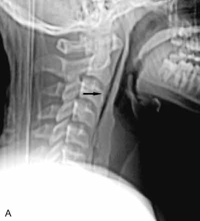
You order radiographs of the soft tissues of the neck.What abnormality is evident on these films, and how willyou proceed to determine its cause?

Figure A

Figure B
2. Throat pain in a catcher hit with a bat: The lateralradiograph reveals a large, linear collection of gas within theretropharyngeal soft tissues (A, arrow). There are 4 possibleexplanations for gas in a location in which it does notnormally occur:
- Communication with a structure that normally containsgas. In this case, the adjacent trachea, the oral cavity, andthe esophagus fit that description.
- Penetrating injuries, as from a gunshot or needle (iatrogenicinjury). This explanation is usually apparent fromthe history.
- Infection with gas-forming organisms. The history andclinical findings will point to this cause.
- Caisson disease, in which a person is subjected to arapid and marked reduction in atmospheric pressure andthe nitrogen in the blood comes out of solution. This typicallyoccurs in rapid ascents from scuba diving.

Figure C

Figure D
In this patient, a laryngeal injury is suspected.You order a CT scan of the neck to further investigatethis possibility. An axial image filmed in bone windowsat the level of the thyroid (B) and an axial image filmedin soft tissue windows at the level of the mandible (C)confirm the finding of retropharyngeal gas (arrows).However, no clearly defined track of gas extends fromthe esophagus or trachea. There is also no clear evidenceof hematoma.
An otolaryngologist performs a nasopharyngoscopy.The results are normal, except for mild edema in the postcricoidregion.
You order a Gastrografin swallow. This anteroposteriorprojection of an esophagram at the level of the softtissues of the neck reveals a clear leakage of contrastmaterial from the cervical esophagus at approximatelythe level of the C6 vertebral body (D, arrow).
This case underscores the importance of persistencein the search for a cause of gas in an unusual locationin a patient who has sustained trauma. A clear causemust be identified so that appropriatetherapy may be instituted.
This patient underwent a neckexploration and surgical repair ofthe tear in his cervical esophagus.A Penrose drain was placed in theretropharyngeal space. His postoperativecourse was uneventful,and a second Gastrografin study 3days later showed no evidence ofleakage.
