- Clinical Technology
- Adult Immunization
- Hepatology
- Pediatric Immunization
- Screening
- Psychiatry
- Allergy
- Women's Health
- Cardiology
- Pediatrics
- Dermatology
- Endocrinology
- Pain Management
- Gastroenterology
- Infectious Disease
- Obesity Medicine
- Rheumatology
- Nephrology
- Neurology
- Pulmonology
Onychomycosis
The prevalence of onychomycosis increases with age; it is less than 1% in persons younger than 19 years and rises to about 18% in those who are 60 to 79 years. The infection is more common in men than in women. Among the predisposing factors are diabetes mellitus, psoriasis, a family history of onychomycosis, use of immunosuppressive drugs, and peripheral vascular disease.
Cutaneous CandidiasisOral CandidiasisPerlche Erosio Interdigitalis BlastomyceticaOnychomycosisTinea PedisTinea CorporisTinea ManuumFungal Folliculitis
Onychomycosis
The prevalence of onychomycosis increases with age; it is less than 1% in persons younger than 19 years and rises to about 18% in those who are 60 to 79 years. The infection is more common in men than in women. Among the predisposing factors are diabetes mellitus, psoriasis, a family history of onychomycosis, use of immunosuppressive drugs, and peripheral vascular disease.

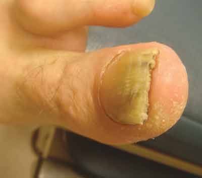
Figure 1 –
This thickened, friable, discolored toenail with subungual hyperkeratosis is characteristic of distal subungual onychomycosis.

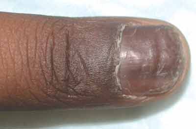
Figure 2 –
Distal subungual onychomycosis can be pigmented.
Onychomycosis is more than a cosmetic problem; it can cause pain and limited mobility. Moreover, onychomycosis and tinea pedis are thought to be risk factors for recurrent cellulitis because they provide a portal of entry for bacterial pathogens. In persons with diabetes, onychomycosis can lead to secondary bacterial infections that can result in foot ulcers, recurrent cellulitis, erysipelas, and gangrene.
Dermatophytes (most commonly, Tinea rubrum) cause onychomycosis, or tinea unguium. Candida and non-dermatophyte molds are rarely implicated as causative agents.
There are 4 types of onychomycosis: distal subungual onychomycosis, proximal subungual onchomycosis, white superficial onychomycosis, and candidal onychomycosis.
Distal subungual onychomycosis, which manifests as thickened and friable nails with associated discoloration and subungual hyperkeratosis, is the most prevalent type; it accounts for 75% to 85% of cases (Figure 1). Sometimes this disorder is pigmented (Figure 2).
The most sensitive and specific way to diagnose onychomycosis is by periodic acid-Schiff (PAS) staining of a nail clipping. A definitive diagnosis is useful because psoriasis, lichen planus, and eczema can cause onychodystrophy that resembles onychomycosis. PAS staining cannot distinguish between the different types of dermatophytes; however, this distinction has little bearing on treatment.
Onychomycosis can be treated with oral terbinafine (250 mg/d for 3 months), itraconazole (400 mg/d for 1 out of every 4 weeks, for 12 weeks), or fluconazole (400 mg weekly for 48 weeks). Shorter courses of oral antifungal therapy can be used if only fingernail onychomycosis is present, but the exact duration of such therapy has not been determined in large-scale clinical trials. Ciclopirox nail lacquer is another treatment option for onychomycosis. Terbinafine is the most effective agent and has few drug interactions, but it is effective only against dermatophytes; the other agents are also effective against nondermatophytes. Topical application of urea 40% gel or cream, which breaks down keratin and softens the nail plate, may enhance penetration of topical antifungal drugs.
It can take up to 6 months to see the effect of these therapies on toenail onychomycosis. Combinations of treatments (eg, oral and topical or oral/topical and surgical) appear to be associated with enhanced cure rates. Advise patients to disinfect or discard old footwear, keep the feet dry and clean, and continue to use topical antifungal agents.
References:
FOR MORE INFORMATION:
- Brodell RT, Elewski B. Superficial fungal infections. Errors to avoid in diagnosis and treatment. Postgrad Med. 1997;101(4):279-287.
- Loo DS. Onychomycosis in the elderly: drug treatment options. Drugs Aging. 2007;24:293-302.
- Tan JS, Joseph WS. Common fungal infections of the feet in patients with diabetes mellitus. Drugs Aging. 2004;21;101-112.
- Weinberg JM, Scheinfeld NS. Cutaneous infections in the elderly: diagnosis and management. Dermatol Ther. 2003;16:195-205.
- Weinberg JM, Vafaie J, Scheinfeld NS. Skin infections in the elderly. Dermatol Clin. 2004;22:51-61.
