- Clinical Technology
- Adult Immunization
- Hepatology
- Pediatric Immunization
- Screening
- Psychiatry
- Allergy
- Women's Health
- Cardiology
- Pediatrics
- Dermatology
- Endocrinology
- Pain Management
- Gastroenterology
- Infectious Disease
- Obesity Medicine
- Rheumatology
- Nephrology
- Neurology
- Pulmonology
Man With Severe Eye Pain, Impaired Vision, and Purulent Discharge
Photo Finish

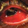
The Case: A 42-year-old man presents with severe pain, decreased vision, photophobia, and purulent discharge in his right eye. Four days earlier, after debris thrown up by a vehicle struck his eye, he noted slight redness, but the other ocular symptoms did not develop until the day before he sought medical attention.
The patient wears contact lenses. He cleans them daily; occasionally, he sleeps in them but denies doing this recently. He has type 2 diabetes mellitus, which he controls with diet, and hypertension, which is poorly controlled because he is noncompliant with his prescribed medications.
Temperature is 37.5°C (99.4°F); blood pressure, 190/120 mm Hg; and heart rate, 90 beats per minute; respiration rate is normal. No proptosis is noted in the right periorbital area, but there is significant chemosis, injection, and purulence. A small amount of pus is also noted in the anterior chamber. The patient can see vague shadows on visual acuity testing of the right eye. His pupils are equal, round, and reactive to light. Slit-lamp examination reveals a central 4-mm ulceration with purulent drainage. A Seidel test for leaking aqueous humor is negative.
What is the most likely cause of this patient's symptoms?
• Corneal ulcer
• Traumatic endophthalmitis
• Conjunctivitis
• Iritis/uveitis
(Answer and discussion on next page)

DISCUSSION: This patient was emergently referred to an ophthalmologist. Culture of a corneal swab grew Pseudomonas aeruginosa, which confirmed the suspected diagnosis of a corneal ulcer. Multiple organisms may cause corneal ulceration, including bacteria (most notably Pseudomonas), fungi, and the protozoa Acanthamoeba. In addition, mycobacteria may be found in patients with corneal grafts.
Corneal ulceration is usually very painful. It typically occurs in contact lens users who have poor hygiene, although all those who wear contact lenses (regardless of hygiene) as well as persons who have diabetes are at heightened risk. This patient's infection was probably secondary to minor trauma from his contact lens use or from the debris sprayed by the vehicle.
The diagnosis of corneal ulcer is based on a thorough history taking and examination. Slit-lamp examination must include a description of the ulcer, size, depth, and location. Intraocular pressure should be measured, and cultures should be obtained from the cornea as well as from the contact lenses, if available. Because ulcerations can jeopardize vision, an urgent ophthalmological consultation is mandatory.
An infectious etiology should be presumed unless the history and examination results suggest another cause. Treatment depends on the severity of the ulceration. Tobramycin or gentamicin drops, alternating with cefazolin or vancomycin drops, are given every hour. Oral ciprofloxacin may be considered in patients with severe ulcers, although oral antibiotics are usually not effective in corneal infections. Adequate analgesia, shielding (not patching), and avoidance of contact lens use until the eye has healed are also recommended. If vision loss or noncompliance is a concern, admit the patient to the hospital. Compliant patients who have a mild ulceration may be discharged with daily follow-up by an ophthalmologist.
This patient received immediate treatment with moxifloxacin (0.5% solution), brimonidine tartrate, and acetazolamide. He was told to apply moxifloxacin, 1 drop every hour for several weeks. Ceftazadime was added for a brief period as was tobramycin ophthalmic ointment (at night). His vision continued to improve. He was scheduled for regular ophthalmological follow-up.
Endophthalmitis-inflammation of the aqueous or vitreous humor-may be classified as either exogenous (caused by a complication of ocular surgery, foreign body, or ocular trauma) or endogenous (a blood-borne infection from a remote source). Symptoms may include eye pain and irritation, vision loss, purulent discharge, and photophobia. Physical examination may reveal chemosis, swelling and erythema of the eyelid, hypopyon, and injected sclera and conjunctiva. The Gram-positive organisms Staphylococcus epidermis,Staphylococcusaureus, and Streptococcus species are most commonly associated with endophthalmitis. However, the Gram-negative bacteria Pseudomonas, Escherichia coli,Enterococcus, and fungi (especially Candida albicans) can also cause this infection. Prompt ophthalmological consultation is needed because timely recognition and treatment can help preserve patients' vision.
Conjunctivitis is one of the most common nontraumatic eye complaints. Although usually self-limited, conjunctivitis can progress to a sight-threatening infection. Symptoms and physical findings vary with the underlying cause (Table).
Treatment consists of antibiotics directed toward the appropriate organisms and symptomatic measures. Consult an ophthalmologist if you suspect anything other than uncomplicated conjunctivitis.
Iritis involves inflammation of the anterior chamber, whereas iridocyclitis affects both the anterior chamber and the ciliary body. Uveitis refers to inflammation of the iris, ciliary body, and choroid. Uveitis may be further classified as either anterior or posterior. Posterior uveitis (choroiditis or retinochoroiditis) is uncommon; it is mostly found in patients with AIDS who have cytomegalovirus retinitis.
Patients with anterior uveitis describe blurry vision, a unilateral painful eye, photophobia, and tearing, whereas those with posterior uveitis complain of occasional pain and photophobia, floaters, and blurry vision. Physical findings of iritis include perilimbal injection (ciliary flush), direct and consensual photophobia as well as papillary miosis. Slit-lamp examination reveals cellular debris (flare) in the otherwise normally clear anterior chamber.
In half of patients with iritis/uveitis, the cause is undetermined. In the remaining half, an underlying systemic disease (such as sarcoid, inflammatory bowel disease, Behet disease, tuberculosis, syphilis, or toxoplasmosis) is responsible; in immunocompromised patients, Cytomegalovirus, herpetic, or candidal infection may be the cause.
Ophthalmological referral is mandatory for patients with iritis/uveitis. Cycloplegics and analgesics should be administered initially. Ocular corticosteroids should not be started until after an ophthalmologist examines the patient.
References:
FOR MORE INFORMATION:
• Brunette D. Ophthalmology. In: Marx J, Hockberger R, Walls R, eds.
Rosen's Emergency Medicine: Concepts and Clinical Practice.
5th ed. St Louis: Mosby; 2002:916-918.
• Eifrig CW, Scott IU, Flynn HW Jr, Miller D. Endophthalmitis caused by
Pseudomonas aeruginosa
.
Ophthalmology.
2003;110:1714-1717.
• Miller JJ, Scott IU, Flynn HW Jr, et al. Endophthalmitis caused by
Streptococcus pneumoniae
.
Am J Ophthalmol.
2004;138:231-236.
• Rhee DJ, Pyfer MF.
The Wills Eye Manual: Office and Emergency Room Diagnosis and Treatment of Eye Disease.
3rd ed. Philadelphia: Lippincott Williams & Wilkins; 1999:72-76, 425-430.
