- Clinical Technology
- Adult Immunization
- Hepatology
- Pediatric Immunization
- Screening
- Psychiatry
- Allergy
- Women's Health
- Cardiology
- Pediatrics
- Dermatology
- Endocrinology
- Pain Management
- Gastroenterology
- Infectious Disease
- Obesity Medicine
- Rheumatology
- Nephrology
- Neurology
- Pulmonology
Lichen Striatus and Relapsing Polychondritis
Dermclinic
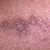
Figure 1
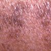
Figure 2
Case 1:
A 35-year-old woman has an asymptomatic, linear, hyperpigmented streak on one leg; it started 2 months earlier on her thigh and slowly progressed down the leg over the next few weeks. The lesions are slightly palpable, with no significant scale, and they do not follow the dermatome.
What does this look like to you?
A.
Inflammatory linear epidermal nevus (LEN).
B.
Linear psoriasis.
C.
Verruca plana (flat warts).
D.
Lichen planus.
E.
Lichen striatus.
(Answer on next page.)

Figure 1

Figure 2
Case 1: Lichen striatus
An irregular band of coalescing hyperpigmented papules that follow the lines of Blaschko on one extremity is a classic presentation of lichen striatus, E. This self-limited eruption usually persists for 1 year before it spontaneously resolves without leaving a mark. Although lichen striatus is more common in children, it can occur in young or older adults. No treatment is necessary; however, some patients may respond to topical corticosteroids or topical tacrolimus.
Inflammatory LEN and linear psoriasis are generally more inflamed and are pruritic. Flat warts usually have a more verrucoid appearance than the lesions seen here and are not as hyperpigmented. Lichen planus typically consists of purple polygonal papules that are pruritic.
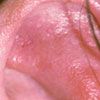
Case 2:
For 2 to 3 days, a 37-year-old woman has had tenderness of 1 ear. It is tender on palpation and when she sleeps on the affected side. There is no history of trauma. A few months earlier, she sought treatment of a similar episode, and cephalexin was prescribed; the tenderness resolved after about 10 days. The patient has been symptom-free until this episode. She is otherwise healthy; she takes an oral contraceptive and ibuprofen as needed for menstrual cramps and headaches.
What do you suspect?
A.
Staphylococcal infection.
B.
Photodrug reaction.
C.
Relapsing polychondritis.
D.
Fixed drug eruption.
E.
Sebopsoriasis.
(Answer on next page.)

Case 2: Relapsing polychondritis
A biopsy revealed inflammation of the cartilage consistent with relapsing polychondritis, C. This recurrent chronic condition is an autoimmune disease directed to type II collagen. Other affected areas can include the trachea, eyes, and joints. Systemic corticosteroids effectively treat acute flares, whereas dapsone reduces the frequency of flares. Most patients require the addition of an immunosuppressive agent to their therapeutic regimen.
A staphylococcal infection is a consideration, but culture would rule it out. Photodrug reactions do not spare the earlobe, as occurred in this patient. Fixed drug eruptions have more epidermal involvement, with induration and crusting. Sebopsoriasis is usually pruritic, not tender, and much more scaling would be present.
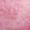
Case 3:
A 49-year-old woman who has a long-standing history of pruritus presents with a new complaint of blisters on her thighs. The itching has been paroxysmal, and there are excoriations on her trunk and extremities. The patient is highly anxious and miserable because of her skin condition. She says the blisters developed during the past few days and are both pruritic and tender. She has been applying triamcinolone cream to the rash for the past several weeks, and she takes oral hydroxyzine to control the pruritus. She denies taking any other medications.
What is the likely cause of the blisters?
A.
Bullous pemphigoid.
B.
Staphylococcal infection.
C.
Bullous impetigo.
D.
Scabies.
E.
Pustular psoriasis.
(Answer on next page.)

Case 3: Linear MRSA infection
A bacterial culture grew methicillin-resistant Staphylococcus aureus (MRSA), B. The patient has lichen simplex chronicus, a neurodermatitis that is often exacerbated by underlying emotional disorders, as in this case. The lesions appeared in a linear pattern because of her scratching.
She was treated with trimethoprim/sulfamethoxazole (Bactrim DS) for the MRSA infection and with topical mupirocin for nasal staphylococcal carrier state. She was also given a selective serotonin reuptake inhibitor to relieve her anxiety pending evaluation by a mental health professional. A new topical skin care regimen with antipruritic moisturizers was recommended. Her symptoms slowly but steadily abated during the following 2 weeks.
Bullous pemphigoid often presents with pruritus before an outbreak. A skin biopsy confirms the diagnosis. Bullous impetigo is usually more honey-crusted than this patient's condition, but the early stages of impetigo may resemble it. Scabies results in excoriated papules that typically occur on the finger webs, axillae, genitalia of men, and nipples of women; secondary infection can occur at any of these sites. Pustular psoriasis usually produces small non-follicular pustules on areas of erythema; affected patients typically have a history of psoriasis.
Case 4:
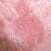
For several weeks, a 58-year-old man has had red, itchy, scaly patches on his face. The rash also involves his scalp and ears but not his trunk or extremities. Switching to a different soap and shampoo has had no effect on the rash.
What is your clinical impression?
A.
Psoriasis.
B.
Seborrheic dermatitis.
C.
Tinea faciei.
D.
Impetigo.
E.
Discoid lupus erythematosus (DLE).
Bonus question: What underlying disease(s) is/are associated with this condition?
(Answers on next page.)

Case 4: Seborrheic dermatitis
This patient has an extensive case of seborrheic dermatitis, B, which affects 2% to 5% of the population. Classic areas of involvement include the scalp, eyelids, eyebrows, ears, nasolabial creases, sternal area, axillae, groin, and gluteal fold. The cause is thought to be related to the Pityrosporum ovale yeast. Treatment is directed toward modifying the inflammatory response with low- potency topical corticosteroids as well as lowering the yeast count with topical antifungal agents.
Psoriasis can be superimposed on seborrhea, but it is generally associated with more inflammation and scaling as well as evidence of psoriasis elsewhere on the body. Tinea faciei can be ruled out with a potassium hydroxide examination. Impetigo typically produces honey-crusted lesions. The patches and plaques caused by DLE are more indurated and less scaly than this patient's rash.
Answer to bonus question: Extensive seborrheic dermatitis is associated with Parkinson disease and AIDS.
FDA Approves Guselkumab for Pediatric Plaque Psoriasis and Active Psoriatic Arthritis
September 30th 2025The FDA has approved guselkumab for children aged 6 years and older with moderate to severe plaque psoriasis or active psoriatic arthritis, making it the first IL-23 inhibitor authorized for pediatric use.
