- Clinical Technology
- Adult Immunization
- Hepatology
- Pediatric Immunization
- Screening
- Psychiatry
- Allergy
- Women's Health
- Cardiology
- Pediatrics
- Dermatology
- Endocrinology
- Pain Management
- Gastroenterology
- Infectious Disease
- Obesity Medicine
- Rheumatology
- Nephrology
- Neurology
- Pulmonology
Kidney Disease: A Straightforward Diagnostic Approach
Chronic kidney disease (CKD) has become a burgeoning epidemic. Patients with various stages of CKD initially seek care from their primary care physician; some of these patients sustain acute, reversible renal injuries as well.
ABSTRACT: Certain indicators derived from noninvasive testing can categorize kidney diseases into clinically relevant groups. The results of select tests- estimates of glomerular filtration rate (GFR), renal ultrasound scans, and urinalysis, in conjunction with urinary protein measurements-can be used to determine whether renal injury is acute or chronic, glomerular or interstitial, in a systematic manner. First, GFR can be estimated and kidney disease staged if it is chronic (of greater than 3 months’ duration). Then, ultrasound examination can further determine the duration of disease, the presence of obstruction, and the possibility of a solitary functioning kidney or other anatomic abnormalities. Finally, urinalysis and spot urine evaluations for both protein and creatinine can lead to determinations that implicate the glomerulus or interstitium as the origin of renal injury.
Key words: chronic kidney disease, renal disease
Chronic kidney disease (CKD) has become a burgeoning epidemic. Patients with various stages of CKD initially seek care from their primary care physician; some of these patients sustain acute, reversible renal injuries as well.
If the renal injury is to be reversed, or at least slowed in its progression to dialysis dependence, its presence and cause must be ascertained. Timely nephrology referral and appropriate treatment-such as blood pressure control with angiotensin-converting enzyme (ACE) inhibitors or angiotensin receptor blockers (ARBs)-have become commonplace management tools in primary care practice.
In this article, we present a diagnostic algorithm designed to streamline the approach to kidney disease for primary care practitioners. The Algorithm relies on select noninvasive tests-estimates of glomerular filtration rate (GFR), renal ultrasound scans, and urinalysis, in conjunction with urinary protein measurements-to categorize disease processes and to determine whether renal injury is acute or chronic, glomerular or interstitial, in a systematic manner. Despite its simplicity, this approach is accurate, inexpensive, and efficient. We will also apply the algorithm to real-world cases representative of those seen in primary care practice.
NOMENCLATURE AND ETIOLOGY OF RENAL DISEASE: SOURCES OF CONFUSION
The complex vocabulary surrounding kidney disease has been in constant flux and, as a result, multiple names have been used for the same disease. Thus, it should come as no surprise that unnecessary confusion has been engendered. For example, during the past generation, minimal-change disease, lipoid nephrosis, and nil disease have been used interchangeably to describe the same nephrotic lesion. Another example is rapidly progressive glomerulonephritis (RPGN) and crescentic glomerulonephritis. Both names describe the same pathology comprising glomerular crescents on renal biopsy and a rapidly progressive decline in renal function.
To further complicate the issue, the etiology of RPGN has exploded into a confusing laundry list of diseases that run the gamut from Wegener granulomatosis to Henoch-Schönlein purpura. If competing names are not enough, some diseases, such as diabetic renal failure, can lead to a chronic vascular renal injury (nephrosclerosis) without significant proteinuria, a glomerular injury with nephrotic range proteinuria, or even acute kidney failure as a consequence of contrast media administration.
The confusion has caused some physicians to avoid renal diagnostic evaluations completely. Now that kidney disease has become a common problem-presenting to primary care at a stage at which it is still treatable-this avoidance has become unacceptable. If a decline in the number of persons who progress to dialysis dependence is to be realized, or at least a slowing of progression, primary care has to become the critical diagnostic and therapeutic interface for kidney disease.
PRIMARY CARE: THE FRONT LINE IN MANAGING RENAL DISEASE
Typically, physicians are confronted with evidence of kidney disease through abnormal urinalysis results or an elevated serum creatinine level, frequently in patients for whom past information is meager. The abnormality is often “quantitated” either in terms of a decreased GFR or “dipstick 1-4+” proteinuria.
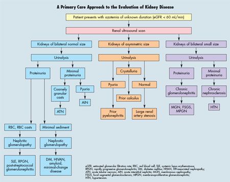
The initial steps in diagnosis are critical: to establish through imaging whether deficits in kidney function, or GFR, are acute or chronic. The latter is defined as decreased GFR or structural kidney damage of greater than 3 months’ duration. Then, judiciously chosen tests guide a diagnostic scheme that when followed systematically implicates the site of renal injury through a reliance on measurement of urinary protein and sediment (see Algorithm). As Cases 1 through 4 will illustrate, a structural examination of the kidneys with ultrasonography, paired with a noninvasive “biopsy” of the urine through urinalysis, serves as an effective primary care entry into the diagnostic process.
Ultrasonography remains the screening test of choice for pinpointing urinary tract obstruction as well as differentiating acute from chronic renal disease on the basis of kidney size. Additional information obtained from the ultrasound, such as the discovery of cysts in patients with autosomal dominant polycystic kidney disease (ADPKD) or the detection of a renal size disparity suggestive of renal artery stenosis, is provided without further cost or administration of contrast.
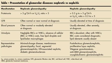
The urinalysis forms the basis both for identifying patients with glomerular disease and for broadly categorizing such diseases as either “nephritic” or “nephrotic.” Since only primary care is designed to span multiple organ systems simultaneously, the diagnosis of serious systemic diseases with renal manifestations-for instance kidney, electrolyte (calcium), bone, and blood abnormalities accompanying multiple myeloma-can be quickly “pieced together” through the renal algorithm.
(Article continues on next page)
KEYS TO EVALUATION OF DECREASED RENAL FUNCTIONFunctional evaluation. Renal function is defined by GFR or by the closely related value of creatinine clearance. An elevation in serum creatinine level reflects a reduction in GFR and can be used to approximate GFR. In lieu of the traditional, but cumbersome, 24-hour urine collection used to measure creatinine clearance as a surrogate for GFR, the preferred method is to apply validated formulas that incorporate serum creatinine levels and select demographic data. As a result, most laboratories now report an estimated GFR as calculated from the Modification of Diet in Renal Disease (MDRD) or other formulas.1-3
Furthermore, CKD is presently categorized by stages on the basis of estimated GFR. Documented kidney disease of 3 months’ or greater duration is staged as follows:
• CKD-1 (GFR, 90 mL/min or higher).
• CKD-2 (GFR, 60 to 89 mL/min).
• CKD-3 (GFR, 30 to 59 mL/min).
• CKD-4 (GFR, 16 to 29 mL/min).
• CKD-5 (GFR, 0 to 15 mL/min).
The stages are relevant in that stage CKD-3 heralds referral to a nephrologist and stage CKD-5 is consistent with dialysis dependence or transplant.
Anatomical evaluation. The renal ultrasound is the single most valuable determination in assessing the duration of kidney disease (acute or chronic). If the ultrasound scan demonstrates adult kidneys that are small (approximately 8 cm in length or less), chronic renal disease is assumed. In contrast, if the kidneys are larger (generally more than 10 cm in length), the renal injury is more likely acute and therefore potentially reversible. Note that enlargement of the kidneys is also associated with certain chronic conditions, such as diabetes mellitus, amyloidosis, and infiltrative cancers.
Acute injury superimposed on chronic disease can be similarly determined. For example, contrast nephropathy might be suspected as superimposed on chronic diabetic nephropathy (stage 3 CKD) if a renal ultrasound scan demonstrates 9-cm kidneys (smaller than expected), the estimated GFR for more than 3 months is 45 mL/min, and a further acute reduction in GFR to less 10 mL/min occurs after contrast administration.
If measurements of the two kidneys on an ultrasound scan are different (disparity of more than 2 cm), the smaller kidney may have been injured in the past by ischemia (renal artery stenosis) or another unilateral pathological process, such as obstruction or infection. In addition, ultrasonography can detect polycystic kidney disease, the most common inherited disease of the kidney; identify residual renal calculi, a potential cause of obstruction; or reveal papillary necrosis, a clue to underlying sickle cell disease or analgesic abuse.
Although obstructive uropathy, or post-renal failure, is not the most common cause of either acute or chronic renal failure, it may be a completely correctable form of kidney disease-but only if discovered early by an ultrasound scan. Therefore, it is good practice to assume that any patient with kidney disease might have an obstruction.
The diagnosis of obstructive uropathy is made when hydronephrosis is demonstrated by an ultrasound scan. An autopsy study in 59,064 persons found that hydronephrosis was present in 3.1%, but in those who died after age 60, in as many as 5.1%.4 Tseng and Stoller4 also observed that 9.5% of elderly patients admitted to the hospital for acute renal failure had post-renal, or obstructive, abnormalities.
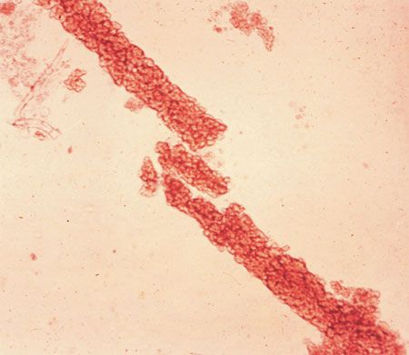
Figure 1 - Red blood cell (RBC) casts and RBCs are visible in the urine of a woman with nephritic glomerular disease (x40). (Courtesy of Dr George Schreiner.)
If hydronephrosis is responsible for a decrease in GFR, it will be bilateral and will usually originate in the lower urinary tract. However, if a solitary functioning kidney has hydronephrosis-meaning that the other kidney is either absent or atrophic-a stone that obstructs the functioning kidney can be the sole cause of acute renal failure. If the stone is discovered, the obstruction can be reversed.
(Article continues on next page)
Pathological evaluation. The urinalysis is actually a surrogate but noninvasive renal “biopsy” that provides clues to important kidney pathology. Critical to the assessment of kidney disease is the presence or absence of proteinuria. A “snapshot” of urinary protein by dipstick must be considered in light of the concentration of the urine specimen in which it is measured; that is, the more concentrated the urine, as reflected by the dipstick specific gravity, the less likely the actual amount of protein is significant.
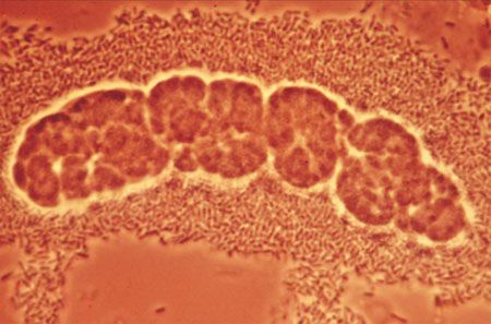
Figure 2 - White blood cell casts are surrounded by numerous bacteria in this urine specimen from a patient with pyelonephritis (x100). (Courtesy of Dr George Schreiner.)
If a dipstick specimen suggests significant proteinuria, a more accurate estimate of urinary protein excretion should be obtained by a quantitative measurement of urinary protein (Uprotein) and urinary creatinine (Ucreatinine) concentrations. This can be accomplished with a spot urine sample. The ratio of Uprotein to Ucreatinine, a dimensionless number, correlates to the grams of protein expected if a simultaneous 24-hour urine collection were performed. However, the spot sample is much easier to obtain. A ratio of 0.3 or less, indicating a protein excretion of 0.3 g or 300 mg/24 h, is considered normal or minimal. A ratio of 3.0 or higher is consistent with “glomerular” range proteinuria, or so-called nephrotic range proteinuria. This ratio can be seen in membranous nephropathy, for instance. Values between 0.3 and 3.0 indicate kidney disease but are not definitive in themselves (based on sensitivity and/or specificity) for a glomerular locus.
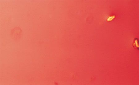
Figure 3 - Calcium oxalate crystals are shown in this urine specimen (x40). (Courtesy of Dr George Schreiner.)
The urinary sediment-cells, casts, and crystals-can also point to specific renal pathology. The presence of red blood cells (RBCs), especially dysmorphic ones, and RBC casts (Figure 1) suggests a “nephritic” glomerulopathy (Table). An example of a nephritic process might be vasculitis with glomerular involvement as may occur in RPGN. Abundant white blood cells (WBCs) in urinary sediments (Figure 2) can point to infection (such as pyelonephritis) or drug-induced interstitial nephritis. Crystals (eg, oxalate [Figure 3], cystine [Figure 4], uric acid [Figure 5]) can suggest stones in the setting of pain or obstruction found on an ultrasound scan.
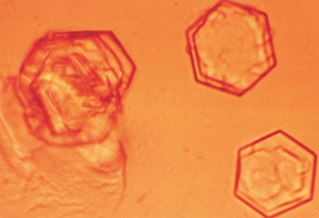
Figure 4 - Cystine crystals are evident in this urine specimen (x100). (Courtesy of Dr George Schreiner.)

Figure 5 - Uric acid crystals (arrow) are shown in this urine specimen (x40). (Courtesy of Dr George Schreiner.)
(Article continues on next page)
CASE 1 - MAN WITH RESISTANT HYPERTENSION AND FAMILY HISTORY OF RENAL DISEASEClinical background. A 35-year-old man presents for primary care establishment after relocating for an employment opportunity. His medical records reveal that he has been treated for hypertension and a slightly elevated creatinine level (1.7 mg/dL 3 months earlier; estimated GFR, 62 mL/min). His blood pressure has been elevated since he was 23 years old (then 150/98 mm Hg) and has not been at target on a regimen of an ACE inhibitor (lisinopril, 10 mg/d) and a diuretic (hydrochlorothiazide, 12.5 mg qd). He has been otherwise healthy and up-to-date on his vaccinations.
Some of his family members have had difficult-to-control hypertension. His father and paternal uncle both died in their sixties from complications of hypertension: a stroke in his father and end-stage renal disease in his uncle who died of a myocardial infarction while receiving dialysis. He does not know what specific problem led to his uncle’s dialysis dependence.
The patient has not had any operations or hospitalizations, and he has no known drug allergies. A fasting lipid panel 3 years earlier revealed a total cholesterol level of 246 mg/dL, but he declined medical treatment. He started an exercise program because his body mass index (BMI) was 28 kg/m2.
His blood pressure is 148/100 mm Hg, and his BMI is now 24 kg/m2. Aside from trace pedal edema, physical findings are normal. ECG findings are consistent with left ventricular hypertrophy and “strain pattern.”
Laboratory tests demonstrate the following values: blood urea nitrogen (BUN), 19 mg/dL; serum creatinine, 1.8 mg/dL; and fasting total cholesterol, 280 mg/dL. On the basis of this laboratory report, his estimated GFR is 59 mL/min (from the MDRD equation), which-together with his data from 6 months earlier-classifies him as having stage 3 CKD (ie, present for longer than 3 months and GFR is less than 60 mL/min but greater than 30 mL/min). How would you proceed?
Applying the algorithm. Since you have evidence of CKD from serial MDRD estimates, you order an ultrasound and urinalysis according to the Algorithm. The ultrasound scan is shown in Figure 6. The urinalysis reveals 1+ urinary dipstick protein, and no sediment is appreciated. A spot Uprotein to Ucreatinine ratio is less than 0.3. Since ADPKD is not a glomerular disease, this result is not unexpected. By applying the results of serial GFR estimates, an ultrasound scan, urinalysis, and spot urinary protein determination, a diagnosis of CKD-3 caused by ADPKD can be entertained.
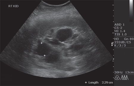
Figure 6 - Cysts are evident on this renal ultrasound scan of a patient with autosomal dominant polycystic kidney disease. The white crosses mark one cyst among others.
In addition to nephrology referral, appropriate primary care management strategies can be implemented.5,6 Blood pressure control (goal, 130/80 mm Hg) with agents that alleviate left ventricular hypertrophy (ACE inhibitors or ARBs) will be the cornerstone of therapy. Since the patient’s blood pressure was not at goal, you add amlodipine to his previous 2-drug regimen. Because cardiac disease is more common in patients with CKD, cholesterol control is also essential. You address his low-density lipoprotein target on the basis of his risk factors after his recent fasting results return.
(Article continues on next page)
CASE 2 - MAN WITH RESISTANT HYPERTENSION AND PROTEINURIAClinical background. Let’s assume that the man from Case 1 presents to your office with the same background history, including onset of hypertension at 23 years of age, accompanied by a reduced GFR for longer than 3 months. The history could also raise suspicion of a chronic glomerular disease that is complicated by secondary hypertension. The hallmark of glomerular diseases is proteinuria, ranging in magnitude from several hundred milligrams to many grams of protein excreted in urine over 24 hours. Initial investigation would again include an anatomic study (an ultrasound scan) complemented by routine urinalysis and spot urinary protein values.
This time, the ultrasound scan demonstrates a 9.8-cm left kidney and a 10.1-cm right kidney. No urinary tract obstruction, cysts, or other anatomic abnormalities are found. The report suggests “medical renal disease.”
The urinary dipstick protein is read as 3+ at a urine specific gravity of 1.010. A spot sample is sent to the laboratory: Uprotein concentration is 134 mg/dL with a Ucreatinine concentration of 42 mg/dL, a ratio of 3.2. Since a ratio of greater than 3 is consistent with glomerular and nephrotic range proteinuria, this spot sample would be consistent with glomerular disease. However, at this juncture, the exact glomerular lesion is unclear. The next steps can add more precision.
Narrowing the differential. The urinalysis can also discriminate between 2 major categories of glomerular disease-nephrotic and nephritic (see Table). This distinction helps differentiate glomerular diseases that cause nephrotic syndrome (membranous nephropathy, focal glomerulosclerosis, HIV-associated nephropathy [HIVAN], for example) from those that are nephritic (hepatitis C renal involvement with cryoglobulins and a membranoproliferative biopsy).
Nephrotic syndrome is defined as urinary protein excretion of more than 3 g/d (as estimated from a spot sample), hypoalbuminemia, edema, and hypercholesterolemia. Nephrotic glomerulopathies have few formed elements (RBCs, WBCs, cellular casts, etc) in urinary sediment, but instead urine may contain free fat droplets or lipid-laden macrophages from the same pathology that causes hypercholesterolemia.
Conversely, when the glomerular lesion is nephritic, urinary protein content is lower (in the glomerular, but not nephrotic, range) and RBCs (often deformed or “dysmorphic” under phase contrast microscopy), WBCs, and cellular casts (such as RBC casts) are present. Such patients generally have less edema, but more hematuria, and occasionally even gross urinary blood. Microscopic examination of this patient’s urine shows 2 to 4 RBCs per high-power field (HPF), 0 to 3 WBCs per HPF, no cellular casts, and an occasional lipid-filled macrophage.
A clue to the duration, and possibly origin, of a chronic glomerulopathy can be obtained with a renal ultrasound scan. As kidney disease progresses, glomerular diseases lead to scarring, thus diminishing kidney size symmetrically; a notable exception is HIVAN. With this specific lesion, kidney size is preserved as end-stage failure approaches. The renal ultrasound scan in this patient reveals kidneys that are “slightly” decreased in size for age (9.1 cm), but are of “increased echogenicity, consistent with medical renal disease.”
Given this patient’s urinary protein excretion in the nephrotic (glomerular) range, his diminished kidney size, and chronic azotemia, the next step in the evaluation would be to perform a serological survey for systemic diseases associated with nephrotic glomerular pathology. The medical history should be carefully reviewed for evidence of hepatitis or exposures that would put the patient at risk for either hepatitis B virus or hepatitis C virus (HCV) infection. Serology for HBsAg and anti-HCV antibody are appropriate, even in the absence of a documented risk.7,8
The patient should be asked about behaviors that increase the risk of HIV infection and, in concert with current recommendations for screening adult patients, an anti-HIV antibody test ordered when appropriate.9 Similarly, a serological test for syphilis, such as a rapid plasma reagin test, should be done. Although systemic lupus erythematosus (SLE) is rare in a white man without physical evidence of rheumatological disease, an antinuclear antibody titer is often a valuable screening test if this disease is suspected. Serum complement levels are rarely of use unless there is clear evidence of a low-complement systemic disease (SLE, hepatitis C nephritic disease, or endocarditis complicated by glomerular disease).
Nonimmunological glomerular disease should also be suspected under appropriate circumstances. For example, the data collected thus far would be compatible with diabetic nephropathy, and measurement of hemoglobin A1c might be appropriate. If markers for metabolic syndrome accompanied the history, the test should be considered.
A history of bone pain, anemia, or hypercalcemia would mandate serum and urine protein electrophoresis with immunofixation to search for a monoclonal gammopathy resulting from multiple myeloma. Although myeloma can be associated with chronic glomerular (amyloid with nephrotic syndrome) or interstitial (light chain disease) presentations, as well as acute injury from contrast, the additional evidence of a “protein gap” (between total albumin and serum globulins), elevated calcium level, and anemia out of proportion to the renal disease are helpful discriminators.
Outcome of this case. Results of serological screening for this patient were positive for anti-HCV antibody. A more directed history revealed that while in college, he “experimented” with drugs, including by injection, although he stopped after graduation. With this information in hand, a decision can now be made as to whether a percutaneous kidney biopsy is warranted. GI specialty consultation, with consideration of ribavirin and pegylated interferon therapy, should also be considered.
Finally, the same background history could have eventuated in a diagnosis of urinary tract obstruction. If a screening ultrasound scan was diagnostic, that may have also explained his chronic azotemia. Obstruction would not have led to significant proteinuria (it does not injure the glomerulus) and may reverse if relieved after diagnosis.
(Article continues on next page)
CASE 3 - HOSPITALIZED MAN WITH ELEVATED CREATININE LEVELClinical background. A 30-year-old man’s creatinine level has risen since he was admitted to the hospital. Two years earlier, he underwent an open reduction-internal fixation of the right lower tibia for a fracture sustained in a motorcycle accident. He also has chronic hypertension, which has been treated with hydrochlorothiazide, 12.5 mg/d, for 1 year. His blood pressure is usually in the 135 to 140/90 to 98 mm Hg range, and his creatinine level was 1.2 mg/dL 3 months earlier. This is consistent with an MDRD GFR estimate of 90 mL/min. Previous urinalysis results were normal.
He has been otherwise healthy and denies a family history of hypertension, diabetes, or kidney disease. However, he recently had fever and chills accompanied by worsening right ankle swelling, erythema, and pain of 2 weeks’ duration. He took ibuprofen, 650 mg 3 times a day, for pain. After he was hospitalized, an MRI scan showed osteomyelitis proximate to the right ankle hardware.
His BUN and creatinine levels are now 19 mg/dL and 1.8 mg/dL, respectively, with a GFR of 62 mL/min. Blood pressure is 145/90 mm Hg (no orthostatic changes are noted); temperature is 37.9°C (100.3°F). Mucous membranes are moist, and skin turgor is good. Findings from the remainder of the examination are unremarkable except for a 3 x 4-cm area of erythema, warmth, and tender swelling over the right lateral ankle. The patient appears to have acute renal failure.
Applying the algorithm. An ultrasound scan demonstrates kidneys that are 11.7 and 12.3 cm in length, without obstruction. Because his history and ultrasound results (normal kidney size) are both consistent with an acute renal insult, the next step is to compare previous and current urinalyses. Results reveal 1+ dipstick protein level, and microscopic examination shows WBC casts and eosinophils detected with a specially ordered Hansel stain (better for finding eosinophils than a Wright stain). The spot Uprotein to Ucreatinine ratio is less than 0.3. These results, accompanied by his clinical history, suggest NSAID-induced interstitial nephritis.
Interstitial renal injuries do not usually cause glomerular range proteinuria. The next step would be discontinuation of the offending NSAID, dosing antibiotics according to a decreased GFR for osteomyelitis, and serial kidney function follow-up. Appropriate attention to electrolytes (sodium and potassium especially) is necessary. Renal biopsy can provide a definitive diagnosis but may not be necessary, especially if kidney function is restored after the NSAID is discontinued. The same can be said about the initiation of corticosteroid therapy.
Acute interstitial nephritis (AIN) can be caused by either medications or infection. It may be implicated in as many as 15% of patients hospitalized for acute renal failure.10 Approximately one-third of cases of drug-related AIN are caused by antibiotics.11
The other diagnosis frequently encountered in inpatients who experience sudden renal failure is acute tubular necrosis (ATN), sometimes called acute renal failure (ARF).12 This entity is responsible for the epidemic of contrast nephropathy.13,14 According to the Algorithm, it would resemble AIN on ultrasound, since it is acute and is accompanied by minimal, nonglomerular urinary protein excretion. Both AIN and ATN affect tubules and surrounding interstitial, but not glomerular, tissue. However, urinary sediment in patients with ATN contains coarse granular casts. Both disorders may require dialysis and definitely warrant renal consultation. In addition to contrast, myoglobinuria from rhabdomyolysis is another cause of ATN or ARF.
(Article continues on next page)
CASE 4 - HYPERTENSIVE WOMAN WITH GROSS HEMATURIA
Clinical background. A woman in her seventh decade presents with gross hematuria.15 She has hypertension (initial blood pressure, 150/98 mm Hg) that is controlled with monotherapy (hydrochlorothiazide, 25 mg/d) and weight loss. Current blood pressure is 130/80 mm Hg. She takes no other prescription or over-the-counter medications. Physical findings are otherwise normal.
Her creatinine level is 1.8 mg/dL, which is consistent with an estimated GFR of 50 mL/min. A GFR from 8 months earlier was 55 mL/min, which suggests CKD-3 of undetermined origin.
Applying the algorithm. An ultrasound scan demonstrates a right renal mass (right kidney, 11.0 cm) and a left kidney of 8.9 cm. Urinalysis results reveal a protein to creatinine ratio of less than 0.3. The sediment has numerous RBCs but no casts. Suspecting renal cell cancer, you obtain a urology consult. The consultant orders a CT scan that is interpreted as follows: “a solid right renal mass (3 x 3.5 cm) that does not appear to have invaded Gerota fascia.” No adenopathy or other organ abnormalities are appreciated. The left kidney is described as “unremarkable.” The patient undergoes nephrectomy.
The nephrectomy and postoperative course are uncomplicated. However, 3 days after surgery, the patient has sudden anuria and a rising creatinine level. There is no evidence of infection; blood loss; hypotension; or use of nephrotoxic medications, including contrast. No obstruction is found on a repeated ultrasound scan of the left kidney. Urinalysis again shows no significant protein or sediment.
This clinical picture is consistent with acute superimposed on chronic (CKD-3) renal failure; prior evaluation suggests that both the previous and recent renal injuries are nonglomerular (low urinary protein levels in spot samples) without obstruction. However, preoperative ultrasound imaging demonstrated a 2.1- cm disparity in the size of the kidneys. Since the typical culprits for ATN (contrast, hypotension, sepsis, nephrotoxins) and AIN (medications, infection) are absent, what might explain the sudden acute portion of the renal failure? The remaining, solitary kidney after nephrectomy was small. Emergency angiography demonstrated complete occlusion of the left renal artery. Emergent revascularization restored renal function after 1 week of dialysis dependence. The patient’s GFR returned to near baseline-47 mL/min. The chronic component of CKD-3 was presumed secondary to renal vascular disease and nephrosclerosis.
LESSONS LEARNED FROM THE CASE STUDIES
Despite the apparent complexity of contemporary renal diseases, certain indicators derived from noninvasive testing can categorize these diseases into clinically relevant groups. First, GFR can be estimated and kidney disease staged if it is chronic (of greater than 3 months’ duration). Then, ultrasound examination can further determine the duration of kidney disease (is kidney size consistent with acute [larger] or chronic [smaller] renal injury?), the presence or absence of obstruction (hydronephrosis), and the possibility of a solitary functioning kidney (a disparity in kidney size) or other anatomic abnormalities (eg, ADPKD). Finally, urinalysis and spot urine evaluations for both protein and creatinine can lead to determinations (a protein to creatinine ratio of greater than 0.3) that implicate the glomerulus or interstitium (lower protein content) as the origin of renal injury. Glomerular diseases can be further subdivided as “nephrotic” or “nephritic” and associated with systemic diseases through pertinent serum values (eg, a positive anti-HCV antibody test that points to hepatitis C as the cause of nephritic glomerular disease).
Although the Algorithm may require supplementation through renal biopsy (for specific glomerular diseases or AIN) and expert consultation (for CKD-3 or as in Case 4), it still relies on the acumen and management skills integral to primary care practice.
References:
REFERENCES:1. Coresh J, Auguste P. Reliability of GFR formulas based on serum creatinine, with special reference to the MDRD Study equation. Scand J Clin Lab Invest Suppl. 2008;241:30-38.
2. Soares AA, Eyff TF, Ritter L, et al. Glomerular filtration rate measurement and prediction equations. Clin Chem Lab Med. 2009;47:1023-1032.
3. Cheung CK, Bhandari S. Perspectives on eGFR reporting from the interface between primary and secondary care. Clin J Am Soc Nephrol. 2009;4:258-260.
4. Tseng TY, Stoller ML. Obstructive uropathy. Clin Geriatr Med. 2009;25:437-443.
5. Bennett WM. Autosomal dominant polycystic kidney disease: 2009 update for internists. Korean J Intern Med. 2009;24:165-168.
6. Schrier RW. Optimal care of autosomal dominant polycystic kidney disease patients. Nephrology (Carlton). 2006;11:124-130.
7. Duffield JS, Qamar A. Advances in the etiology and management of immune-mediated glomerulonephritis. Nephrol Rounds. 2008;6.
8. Baron JP, McDowell LL. A 63-year-old man with hepatitis C and nephrotic syndrome. Am J Kidney Dis. 2007;49:717-720.
9. Winston JA. HIV and CKD epidemiology. Adv Chronic Kidney Dis. 2010;17:19-25.
10. Michel DM, Kelly CJ. Acute interstitial nephritis. J Am Soc Nephrol. 1998;9:505-515.
11. Baker RJ, Pussey CD. The changing profile of acute tubulointerstitial nephritis. Nephrol Dial Transplant. 2004;19:8-11.
12. Fry AC, Farrington K. Management of acute renal failure. Postgrad Med J. 2006;82:106-116.
13. Solomon R. Contrast-induced acute kidney injury (CIAKI). Radiol Clin North Am. 2009;47: 783-788, v.
14. Abu Jawdeh BG, Kanso AA, Schelling JR. Evidence-based approach for prevention of radiocontrast-induced nephropathy. J Hosp Med. 2009;4: 500-506.
15. Roche Z, Rutecki G, Cox J, Whittier FC. Reversible acute renal failure as an atypical presentation of ischemic nephropathy. Am J Kidney Dis. 1993;22:662-667.
The authors report that they have no relevant financial relationships to disclose.
Obesity Linked to Faster Alzheimer Disease Progression in Longitudinal Blood Biomarker Analysis
December 2nd 2025Biomarker trajectories over 5 years in study participants with AD show steeper rises in pTau217, NfL, and amyloid burden among those with obesity, highlighting risk factor relevance.
