- Clinical Technology
- Adult Immunization
- Hepatology
- Pediatric Immunization
- Screening
- Psychiatry
- Allergy
- Women's Health
- Cardiology
- Pediatrics
- Dermatology
- Endocrinology
- Pain Management
- Gastroenterology
- Infectious Disease
- Obesity Medicine
- Rheumatology
- Nephrology
- Neurology
- Pulmonology
Hyperpigmented Nails: Benign Cause-Or Not?
A 75-year-old man is concerned about the discoloration of his fingernails: each nail has dark bands with a clearing in the middle.
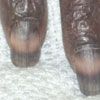
Case 1: Transverse Hyperpigmentation
A 75-year-old man is concerned about the discoloration of his fingernails: each nail has dark bands with a clearing in the middle.
The patient has a history of heart failure, hypothyroidism, hypertension, type 1 diabetes mellitus, proliferative retinopathy, and cataract. Myeloproliferative disorder was diagnosed a year earlier by bone marrow biopsy. He initially was treated with anagrelide; this was later switched to hydroxyurea. A few months before presentation, he temporarily stopped taking the hydroxyurea.
What is the most likely cause of these nail changes?
(Answer on next page.)

Case 1: Reaction to hydroxyurea
This patient’s nail dyschromia was most likely caused by hydroxyurea, which produces hyperpigmentation in about 10% of persons who take the medication, especially those who are dark-skinned.1 The clearing of the middle portion of the fingernails probably correlates to the patient's temporary discontinuation of hydroxyurea a few months earlier.
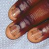
Figure 1
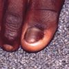
Figure 2
Case 2: Longitudinal Hyperpigmentation
Brown pigmentation of both great toenails and multiple fingernails of a 14-year-old African American boy is noted incidentally during a workup for a fractured thumb. The patient states that the discoloration has been present for several years. He associated the onset of the pigmentation with nail biting, which he did “almost back to the cuticle.”
The patient reports no history of nail infection or trauma-other than the nail biting-and no family history of pigmented nails. He has no prior medical disorders and is not taking any medications.
What is your clinical impression?
(Answer on next page.)

Figure 1

Figure 2
Case 2: Normal finding
Longitudinal brown bands on the nails are a normal finding in more than 90% of African Americans.1 Other causes of brown nails include antimalarial drugs, cancer chemotherapeutic agents, hyperbilirubinemia, junctional nevi, malnutrition, melanocyte-stimulating hormone oversecretion, melanoma (Hutchinson sign), and the use of photographic developer.1
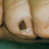
Case 3: Subungual Pigmented Streak
A routine examination of a 54-year-old man reveals an isolated 3-mm dark brown streak on the fourth toe. The patient does not recall any trauma to that foot and was unaware of the streak until it was pointed out to him during the examination. There is no pigmentation of the adjacent skin.
What are you looking at here?
(Answer on next page.)

Case 3: Subungual melanoma
Subungual melanomas account for about 2% of all melanomas; they usually involve the great toe or the thumb. The clinical presentation is a pigmented streak in the nail plate; later, there may be an extension of the pigmentation to the surrounding skin of the finger or toe. The diagnosis is made by punch or excisional biopsy of the nail matrix. Biopsy of the nail plate is not sufficient to make a diagnosis. A high index of suspicion for melanoma is warranted in the case of an isolated subungual pigmented streak in an adult.
Case 4: Hyperpigmented Nail Plate
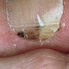
A 45-year-old woman who has had several melanomas removed is concerned about the hyperpigmentation beneath her toenail. Some distal onycholysis is also present. She is not aware of any injury to the toe.
How would you proceed?
(Answer on next page.)

Case 4: Subungual hematoma
Because of the patient’s history of melanoma and the fact that she could not recall injuring the toe, a partial nail evulsion was performed. Fortunately, the only finding was dried blood.
Hyperpigmentation of the nail plate, especially in a patient with a clear history of trauma, is almost always caused by a subungual hemorrhage. If, however, such a history is not forthcoming, biopsy or surgical exploration is strongly recommended. Untreated subungual melanomas can have disastrous consequences.
References:
REFERENCES:1. Hernández-Martin A, Ros-Forteza S, de Unamuno P. Longitudinal, transverse, and diffuse nail hyperpigmentation induced by hydroxyurea. J Am Acad Dermatol. 1999;41:333-334.
2. Habif TP. Clinical Dermatology: A Color Guide to Diagnosis and Therapy. 4th ed. St Louis: Mosby; 2004:886-887.
