- Clinical Technology
- Adult Immunization
- Hepatology
- Pediatric Immunization
- Screening
- Psychiatry
- Allergy
- Women's Health
- Cardiology
- Pediatrics
- Dermatology
- Endocrinology
- Pain Management
- Gastroenterology
- Infectious Disease
- Obesity Medicine
- Rheumatology
- Nephrology
- Neurology
- Pulmonology
An HIV-Infected Woman With Porphyria Cutanea Tarda
A 37-year-old HIV-infected white woman with a CD4+ cell count of 29/µL and an HIV RNA level of 538,000 copies/mL presented with a 2-month history of pruritic blistering eruptions on the dorsal aspects of her hands and feet and hyperpigmentation of the face.
A 37-year-old HIV-infected white woman with a CD4+ cell count of 29/µL and an HIV RNA level of 538,000 copies/mL presented with a 2-month history of pruritic blistering eruptions on the dorsal aspects of her hands and feet and hyperpigmentation of the face. The patient had no associated abdominal pain, neurological symptoms, urine discoloration, or light sensitivity. Her medical history included Pneumocystis jiroveci pneumonia. There was no family history of porphyria. Present medications included nevirapine, tenofovir, emtricitabine, dapsone, and weekly azithromycin. The patient was allergic to trimethoprim/sulfamethoxazole. She was a smoker but denied alcohol and recreational drug use.
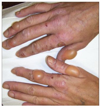
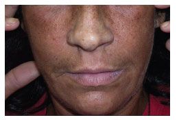
Physical examination revealed multiple tense bullae, vesicles, and crusted lesions on her hands (Figure 1) and feet, as well as hypertrichosis and hyperpigmentation of the face (Figure 2). Other findings were unremarkable.

Figure 1.Bullae, crust, and scars on the dorsum of the hand of an HIV-infected woman with porphyria cutanea tarda.

Figure 2.Hyperpigmentation and hypertrichosis of the face of an HIV-infected woman with porphyria cutanea tarda.
Laboratory test results were as follows: white blood cell count, 3860/µL; hemoglobin, 10.6 g/dL (normal, 12 to 16); ferritin, 1193 ng/mL (normal, 13 to 150); creatinine, 1.1 mg/dL (normal, 0.4 to 1.1); alkaline phosphatase, 251 U/L (normal, 39 to 119); aspartate aminotransferase, 87 U/L (normal, less than 33); alanine aminotransferase, 68 U/L (normal, less than 32); total serum bilirubin, 0.4 mg/dL (normal, 0.0 to 1.0); and γ-glutamyl transpeptidase, 419 U/L (normal, 7 to 32). Results of serological tests for hepatitis A, B, and C viruses were negative. Her 24-hour urinary porphyrin excretion levels were classic for porphyria cutanea tarda: uroporphyrin, 3589 µg (normal, 3 to 25); heptacarboxylporphyrin, 1609 µg (normal, 0 to 7); hexacarboxylporphyrin, 105 µg (normal, 0 to 6); pentacarboxylporphyrin, 91 µg (normal, 0 to 7); coproporphyrin, 39 µg ( normal, 8 to 110); and porphobilinogen, 0.2 mg (normal, 0.0 to 0.5).
Gross pathological examination of a skin biopsy specimen from her right hand showed subepidermal vesiculation, and results of direct immunofluorescence assay showed perivascular IgG, IgA, and fibrinogen.
The patient was treated with low-dose chloroquine. Avoidance of sun exposure was recommended. Her antiretroviral therapy was switched to zidovudine, lamivudine, and ritonavir-boosted atazanavir because of virological and immunological failure despite good adherence to her previous regimen. Two weeks later, the skin lesions resolved without further recurrence.
Porphyria cutanea tarda is a relatively rare skin disorder but still the most common porphyria. It is characterized by blisters, skin fragility in sun-exposed areas, and facial hypertrichosis and hyperpigmentation. Porphyria cutanea tarda is associated with elevated transaminase and γ-glutamyl transpeptidase levels, even when it is not associated with hepatitis C. Porphyria cutanea tarda results from decreased activity of the hepatic enzyme uroporphyrinogen decarboxylase. It is classified into 2 main types: type 1, the sporadic, and most common, form; and type 2, the inherited form. Precipitating factors include alcohol ingestion, estrogen replacement therapy, and hepatitis C.1,2
The incidence of porphyria cutanea tarda is higher in HIV-infected patients than in the general population.3,4 Although hepatitis C is more prevalent in persons with porphyria cutanea tarda and HIV infection can coexist with other risk factors, several reports have shown HIV infection to be an independent risk factor for porphyria cutanea tarda.3-5 The mechanism of HIV infection that causes porphyria cutanea tarda is not fully understood.3-5 The association was initially reported in 1987 by Mansourati and colleagues.4 They reviewed 75 reported cases of HIV infection with porphyria cutanea tarda; almost all (96%) of the patients were male. At the time of diagnosis of porphyria cutanea tarda, the mean age was 36.5 years and the mean CD4+ cell count was 166/µL. Forty-five percent of the patients also had hepatitis C, 55% consumed alcohol heavily, and 89% had abnormal liver function test results. The porphyria cutanea tarda diagnosis preceded the detection of HIV infection in 40% of cases.4 A possible association between the use of protease inhibitors (PIs) and porphyria has been reported in the literature,6 and PI use may be an additional risk factor.
The plasma total porphyrin measurement is a useful initial screening test for porphyria cutanea tarda. Unlike a urine fluorescent screening test for porphyrins, a normal plasma level can exclude the diagnosis of porphyria cutanea tarda. If the plasma total porphyrin level is elevated, urinary and fecal porphyrin levels are needed to confirm the diagnosis, because high plasma total porphyrin levels can be associated with other porphyrias.1,2
The laboratory diagnosis of porphyria cutanea tarda is established by markedly elevated urinary uroporphyrin and heptacarboxylporphyrin levels. Porphyria cutanea tarda is also characterized by increased fecal isocoproporphyrin levels. (No stool testing was done for our patient.) Examination of a skin biopsy specimen using direct immunofluorescence can help differentiate porphyria cutanea tarda from other bullous diseases. When results are positive, this procedure usually reveals deposition of C3 and IgG at the dermal-epidermal junction, which is most apparent in biopsy specimens from sun-exposed areas of persons with active disease.1,2
For persons with porphyria cutanea tarda, avoiding sun exposure and other porphyrinogenic precipitants is important. Phlebotomy and low-dose chloroquine (or hydroxychloroquine) are specific forms of treatment for porphyria cutanea tarda.1,2
The diagnosis of porphyria cutanea tarda should prompt a comprehensive workup for risk factors, including hepatitis C, iron overload, hereditary hemochromatosis genemutation, hepatocellular carcinoma, and HIV infection.
References:
References1. Bickers DR, Frank J. The porphyrias. In: Freedberg IM, Eisen AZ, Wolff K, et al, eds. Fitzpatrick’s Dermatology in General Medicine. 6th ed. New York: McGraw-Hill; 2003:1435-1466.
2. Elder GH. Porphyria cutanea tarda. Semin Liver Dis. 1998;18:67-75.
3. Wissel PS, Sordillo P, Anderson KE, et al. Porphyria cutanea tarda associated with the acquired immune deficiency syndrome. Am J Hematol. 1987;25:107-113.
4. Mansourati F, Stone V, Mayer K. Porphyria cutanea tarda and HIV/AIDS: a review of pathogenesis, clinical manifestations and management. Int J STD AIDS. 1999;10:51-56.
5. Drobacheff C, Derancourt C, Van Landuyt H, et al. Porphyria cutanea tarda associated with human immunodeficiency virus infection. Eur J Dermatol. 1998;8:492-496.
6. Celesia BM, Onorante A, Nunnari G, et al. Porphyria cutanea tarda in an HIV-1 infected patient after initiation of tipranavir/ritonavir: case report. AIDS. 2007;21:1495-1496.
Â
