- Clinical Technology
- Adult Immunization
- Hepatology
- Pediatric Immunization
- Screening
- Psychiatry
- Allergy
- Women's Health
- Cardiology
- Pediatrics
- Dermatology
- Endocrinology
- Pain Management
- Gastroenterology
- Infectious Disease
- Obesity Medicine
- Rheumatology
- Nephrology
- Neurology
- Pulmonology
Histoplasmosis mimicking metastatic carcinoma
The differential diagnosis forendobronchial lesions includesbut is not limited toneoplastic causes, benign tumors,infections, and foreignobjects. We report a case of anunusual cause of endobronchiallesions.
The differential diagnosis for endobronchial lesions includes but is not limited to neoplastic causes, benign tumors, infections, and foreign objects. We report a case of an unusual cause of endobronchial lesions.
The case
A 47-year-old man with a 45-pack-year tobacco history presented to his primary care physician with a 50-lb unintentional weight loss over 3 months, a cough productive of white phlegm, and mouth ulcers. His vital signs were remarkable for the absence of both fever and tachypnea. Physical examination findings were significant for mild cachexia and oral aphthous ulcers.
Laboratory evaluation revealed a normal complete blood cell count but mildly elevated levels of transaminases. A chest radiograph revealed a 2-cm cavitary right upper lobe (RUL) lesion (Figure 1). CT scans of the chest and abdomen revealed the solitary lung lesion, on a background of centrilobular emphysema (Figure 2), and bilateral non-homogeneous adrenal glands, with the left gland appearing larger than the right one (Figure 3). CT scans did not reveal any mediastinal lymphadenopathy or pleural effusions.
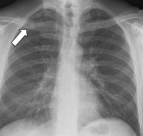
Figure 1 – A cavitary upper lobe mass appears behind the right clavicle in this posteroanterior chest radiograph (arrow).
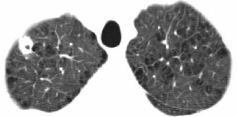
Figure 2 – A 2-cm thick-walled cavitary lesion in the right upper lobe, on a background of emphysema, is revealed in this chest CT scan (5-mm axial cuts, lung window setting).
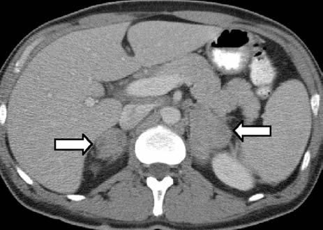
Figure 3 – Bilateral non-homogeneous densities in the adrenal glands can be seen in this abdominal CT scan (5- mm axial cuts) after oral and intravenous administration of contrast (arrows).
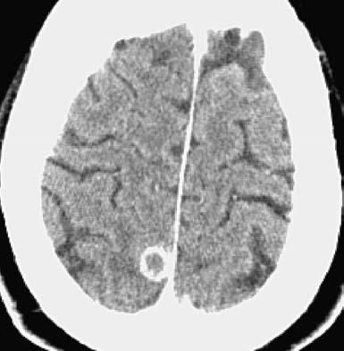
Figure 4 – Ring-enhancing lesions in the right cerebral hemisphere are revealed in this CT scan of the patient's head.
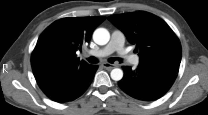
Figure 5 – This CT scan of the chest shows a polypoid endobronchial lesion in the left main-stem bronchus and a calcified periaortic lymph node (5-mm axial cuts, mediastinal window setting).
Before referral to the pulmonary service, the following workup was performed. Initially, CT-guided fine-needle aspiration of the RUL lesion was performed using a 19-gauge needle. Cytological analysis revealed rare atypical cells, suggesting malignancy. Also, pathology revealed necrotizing granulomas, and stains were negative for fungi and mycobacteria. Two subsequent CT-guided left adrenal core biopsies, using a 19-gauge needle, demonstrated necrotic tissue, debris, and a few yeast forms morphologically suggestive of Candida species.
The patient was referred to otolaryngology for a biopsy of the mouth ulcers. The pathology of the left arytenoid and anterior subglottic region revealed ulcers with acute and chronic inflammation, reactive atypia, and yeast-like organisms. After the patient was referred to the oncology clinic with the presumptive diagnosis of metastatic cancer, CT scans revealed numerous small ring-enhancing cortical brain lesions (Figure 4) and a left main-stem endobronchial mass (Figure 5).
On the basis of the chest CT findings, the patient was referred to the pulmonary clinic. The primary team's working diagnosis was metastatic carcinoma; however, our differential diagnosis also included disseminated fungal infection and, less likely, tuberculosis and respiratory papillomatosis. Fungal infection was thought to be less likely after the results of a urinary antigen test for Histoplasma capsulatum, an HIV test, and routine and fungal sputum cultures were reported as negative.
Diagnostic bronchoscopy revealed numerous polypoid lesions lining the trachea and bilateral main-stem bronchi. The lesions were of various sizes, from several millimeters to more than a centimeter in diameter, and were smooth without visible vessels or ulceration (Figure 6). The tracheal lesions were identical in appearance to the polypoid lesions in the main-stem bronchi.
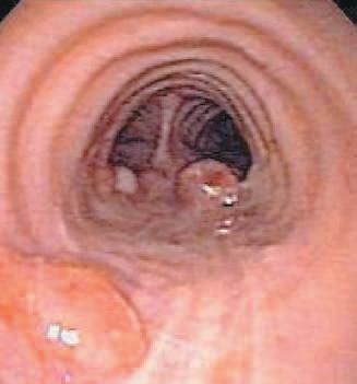
Figure 6 – Bronchoscopy reveals multiple polypoid lesions lining the posterior wall of the trachea.
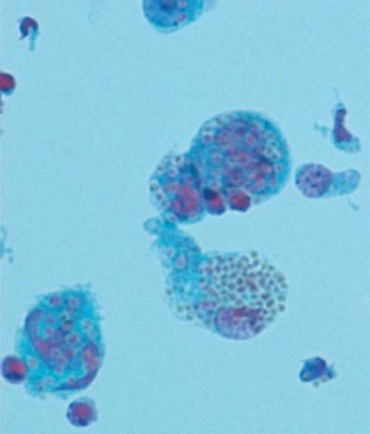
Figure 7 – Alveolar macrophages with engulfed intracellular yeast forms were identified in bronchoalveolar lavage fluid (periodic acid–Schiff stain).
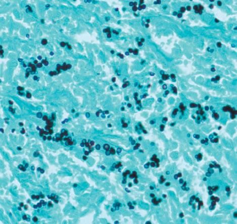
Figure 8 – Biopsy of an endobronchial polyp shows budding yeast forms throughout (methenamine silver stain).
Endobronchial biopsy and bronchoalveolar lavage (BAL) were performed. Cytological analysis of the BAL fluid revealed alveolar macrophages with intracellular yeast forms (Figure 7). The biopsy results revealed multiple small fungal forms with narrow-based budding on silver stain (Figure 8).
After the cytological results were received, the diagnosis of H capsulatum infection was confirmed by bronchoscopy culture results. The patient was admitted and was given amphotericin B for a total of 10 days and then the regimen was switched to oral itraconazole, 400 mg daily.
After the endobronchial lesion biopsy results were reported as histoplasmosis, the other biopsy specimens (lung, adrenal, and arytenoid biopsies) were reviewed again and were found to be consistent with H capsulatum and not Candida.
After a rash developed, itraconazole treatment was switched to voriconazole, 200 mg twice daily, which was continued for 12 months. CT scans of the brain and chest demonstrated regression of all lesions without complete resolution. Clinically, the patient's energy returned, and he regained 25 lb.
The patient's final diagnosis was chronic progressive disseminated histoplasmosis with endobronchial histoplasmosis confirmed by both histology and culture.
Discussion
This case demonstrates 2 important teaching points. First, it is a great example of the clinical findings of chronic progressive disseminated histoplasmosis. Second, and equally important, this case highlights the value of establishing a comprehensive differential diagnosis at the outset. The members of the primary team focused their workup solely on metastatic carcinoma. Had they considered other diagnoses, the "negative biopsies" would have led them to the diagnosis of histoplasmosis much earlier. This "tunnel vision" resulted not only in a delay in arriving at a diagnosis and an effective treatment but also in a number of potentially unnecessary procedures.
To minimize error and unnecessary delays, medical decision making should be a dynamic process wherein one starts with an appropriately wide differential diagnosis, thus avoiding the situation noted here in which data were forced to fit a preconceived-and incorrect-diagnosis.
H capsulatum is a dimorphic fungus endemic to the Ohio and Mississippi river valleys. High concentrations of H capsulatum are found in nitrogen-rich soil, caves with bat guano, and areas containing bird feces. Some activities that can lead to infection include cleaning barns, landscaping, and spelunking.
Inhalation of the organisms is the primary mode of infection. These small organisms are phagocytosed by macrophages in the alveoli and are converted to the yeast form. The yeast forms are typically small (2 to 4 μm) and uninucleated, with single narrow-based buds. The organism lives in the macrophage and is hematogenously disseminated via the reticuloendothelial system. Specific cell-mediated immunity against H capsulatum develops after several weeks, which activates macrophages to the organism.
Our patient presented in a medical facility in Richmond, Virginia, where H capsulatum is not endemic, and he denied any recent travel history, so the initial clinical suspicion for histoplasmosis was low. His definitive source of infection remains unknown. It is postulated that isolated cases in regions where H capsulatum is not endemic represent either geographic microfoci of disease or reactivation of latent disease acquired during previous travel to areas where the fungus is endemic.1
The extent of disease depends on the number of organisms inhaled and the immune response of the host. Most infected patients remain asymptomatic or have acute pulmonary histoplasmosis, which is a mild, self-limited disease that lasts 1 to 3 weeks. The usual symptoms include fever, chills, fatigue, myalgias, dyspnea, nonproductive cough, and anterior chest pain.
Chest radiographs can show patchy airspace consolidations with mediastinal or hilar lymphadenopathy. Without treatment, the pulmonary abnormalities will either clear spontaneously or they will become "walled off" through a granulomatous and fibrotic reaction that can form a solitary pulmonary nodule or histoplasmoma.2 In patients who have underlying chronic obstructive lung disease, it is common for histoplasmomas to cavitate.3,4 Rare complications of pulmonary histoplasmosis include pleural effusions, granulomatous mediastinitis, fibrosing mediastinitis, tracheobronchial stenosis, broncholiths, and endobronchial histoplasmosis.5
Progressive disseminated histoplasmosis (PDH) is a progressive form of the illness with fever and wasting, most often seen with a large inoculum or in the setting of a T-lymphocyte–mediated immunosuppression. Patients with acute PDH have high fevers, dyspnea, nonproductive cough, and diffuse pulmonary infiltrates that can quickly progress to acute respiratory distress syndrome.
Chronic PDH is seen almost exclusively in otherwise healthy middle-aged to elderly men. Patients at increased risk for chronic PDH are those older than 40 years and smokers who have preexisting lung disease.6 Low-grade fever, malaise, weight loss, and night sweats usually occur for months before diagnosis. Many patients with chronic PDH present with laryngeal and oropharyngeal disease.1
A chest radiograph may reveal patchy airspace consolidation in one or more lobes, with associated lymphadenopathy. Laboratory findings include increased erythrocyte sedimentation rate, pancytopenia, and elevated alkaline phosphatase levels.
Our patient had the risk factors, time course, symptoms, laryngeal ulcers, and laboratory findings consistent with chronic PDH, but his endobronchial lesions were extremely atypical for histoplasmosis. The endobronchial lesions were not consistent with broncholiths, since they were not hard, yellowish material with surrounding granulation tissue.7
Regarding the formation of our patient's endobronchial lesions, it is possible that the airways had been seeded via endobronchial spread from his RUL cavitary lesion; however, the pathogenesis of this particular endobronchial manifestation is unknown. His asymptomatic brain lesions were more consistent with metastatic malignancy than with histoplasmosis. The cavitary lung lesions were consistent with a diagnosis of pulmonary histoplasmosis but were also consistent with tuberculosis, nontuberculous mycobacterial infection, neoplasm, and other endemic fungal infections.8 It is not always possible to differentiate between these processes solely on radiographic findings.9
Most commonly reported airway findings in histoplasmosis are normal mucosa and nonspecific mucosal inflammation; there can also be airway narrowing secondary to extrinsic compression from lymphadenopathy. In addition, there have been reports of mucosal ulceration and sloughing and broncholithiasis.
A recent review of cases of endobronchial fungal disease reported in 2005 and cited in Medline database identified 228 cases with only 11 involving histoplasmosis.5 In these 11 cases, the female-to-male ratio was 8:3, and in all 11 cases, the patients were reported to be immunocompetent. The most prominent presenting symptom was hemoptysis related to fibrosing mediastinitis. Bronchoscopic findings varied from hyperemic mucosa to bronchoesophageal fistula, with no reports of endobronchial masses.5
A small case series from Ohio State University described how histoplasmosis may masquerade as a primary endobronchial neoplasm.10 The authors described 4 patients with hemoptysis who underwent bronchoscopy for biopsy of suspected carcinoma but were found to have endobronchial histoplasmosis. Two of these patients presented with single friable endobronchial masses adherent to the mucosa, similar to the lesion in our patient's left main-stem bronchus. On the basis of CT findings, neither these lesions nor our patient's lesions were thought to be calcified lymph nodes. It was postulated that the lesions represented a broncholith that once in contact with the mucosal surface, led to an exuberant inflammatory response with fibrosis and neovascularization.10
There were several differences between the above-described case series and our patient. In 3 of the 4 patients, parenchymal infiltration and fibrosis were found on a CT scan and through histology after resection. Also, these patients presented with single endobronchial masses, while our patient had studding of the trachea and main-stem bronchi with numerous masses.
The diagnosis of histoplasmosis is often made by biopsy of a lesion or by fluid cytology in combination with confirmatory serological studies. Histopathological examination using methenamine silver stain or periodic acid–Schiff stain reveals the small narrow-based budding cells, which may be present intracellularly in macrophages. H capsulatum must be distinguished from other organisms, such as Pneumocystis jiroveci, as well as from other fungi, including Blastomyces dermatitidis, Candida, and Coccidioides immitis. B dermatitidis has broad-based buds, is larger, and has a thick wall; Candida species are rarely found inside macrophages and cause a different clinical picture; and Coccidioides shows spherules and no budding yeast on histological examination. Definitive diagnosis through culture growth of H capsulatum from tissue, sputum, blood, urine, or other body fluid may take several weeks.
Serology is often helpful for preliminary tests while culture or cytological results are awaited. Complement fixation and immunodiffusion tests are available through reference laboratories. Immunodiffusion is more specific than complement fixation (greater than 95% specificity vs 85% to 90%, respectively), while both are only modestly sensitive (75% to 85% sensitivity). 11 These tests are less reliable in immunosuppressed patients. In most patients with chronic PDH, complement fixation antibodies are present.
Enzyme immunoassay (EIA) measuring the cell wall polysaccharide antigen of H capsulatum in the patient's urine and serum can be helpful in acute histoplasmosis, but results are usually negative in patients with chronic pulmonary histoplasmosis. For EIA, a urine sample is more sensitive than a serum sample. Our patient had negative urine and serum EIA results, but serological testing was not done.
Treatment of histoplasmosis depends on the severity of illness.12 Patients with mild to moderate histoplasmosis are treated with an azole antifungal, preferably itraconazole. Those with severe histoplasmosis are given amphotericin B then oral itraconazole after clinical improvement. The length of treatment depends on disease severity. Treatment should continue until there is a complete resolution of symptoms and radiographic infiltrates; this usually takes at least 6 months. In chronic PDH, itraconazole should be continued for 12 to 24 months to prevent the progression of infection.
Conclusion
While it is an extremely unusual presentation of histoplasmosis, this case illustrates the importance of considering disseminated fungal disease in the differential diagnosis of endobronchial lesions and apparent metastatic disease, the need for tissue diagnosis, and the importance of alerting the pathologist to these concerns.
References:
REFERENCES
1. O'Hara CD, Allegretto MW, Taylor GD, Isotalo PA. Epiglottic histoplasmosis presenting in a nonendemic region: a clinical mimic of laryngeal carcinoma. Arch Pathol Lab Med. 2004;128:574-577.
2. Wallace JM. The role of bronchoscopy in pulmonary mycoses. J Bronchol. 2001;8:114-122.
3. Chick EW, Bauman DS. Editorial: Acute cavitary histoplasmosis-fact or fiction? Chest. 1974;65:497.
4. Wheat LJ, Wass J, Norton J, et al. Cavitary histoplasmosis occurring during two large urban outbreaks. Analysis of clinical, epidemiologic, roentgenographic, and laboratory features. Medicine (Baltimore). 1984;63:201-209.
5. Karnak D, Avery RK, Gildea TR, et al. Endobronchial fungal disease: an under-recognized entity. Respiration. 2007;74:88-104.
6. Aziz R, Khan N, Qayum I, Khan AR. Histoplasmosis-case report. J Ayub Med Coll Abbottabad. 2002;14:42-44.
7. Seo JB, Song KS, Lee JS, et al. Broncholithiasis: review of the causes with radiologic-pathologic correlation. Radiographics. 2002;22:S199-S213.
8. Kauffman CA. Histoplasmosis: a clinical and laboratory update. Clin Microbiol Rev. 2007;20:115-132.
9. Goodwin RA Jr, Owens FT, Snell JD, et al. Chronic pulmonary histoplasmosis. Medicine (Baltimore). 1976;55:413-452.
10. Ross P Jr, Magro CM, King MA. Endobronchial histoplasmosis: a masquerade of primary endobronchial neoplasia-a clinical study of four cases. Ann Thorac Surg. 2004;78:277-281.
11. Wheat J, French ML, Kamel S, Tewari RP. Evaluation of cross-reactions in Histoplasma capsulatum serologic tests. J Clin Microbiol. 1986;23:493-499.
12. Wheat LJ, Freifeld AG, Kleiman MB, et al; Infectious Diseases Society of America. Clinical practice guidelines for the management of patients with histoplasmosis: 2007 update by the Infectious Diseases Society of America. Clin Infect Dis. 2007;45:807-825.
