- Clinical Technology
- Adult Immunization
- Hepatology
- Pediatric Immunization
- Screening
- Psychiatry
- Allergy
- Women's Health
- Cardiology
- Pediatrics
- Dermatology
- Endocrinology
- Pain Management
- Gastroenterology
- Infectious Disease
- Obesity Medicine
- Rheumatology
- Nephrology
- Neurology
- Pulmonology
Herpes Zoster: A Photo Essay
Herpes zoster is a reactivation of an infection with the varicella zoster virus, manifests as painful vesicular rash.
These images illustrate a classic rash indicative of herpes zoster. The location of the macular and vesicular lesions suggests involvement of the T4 dermatome.
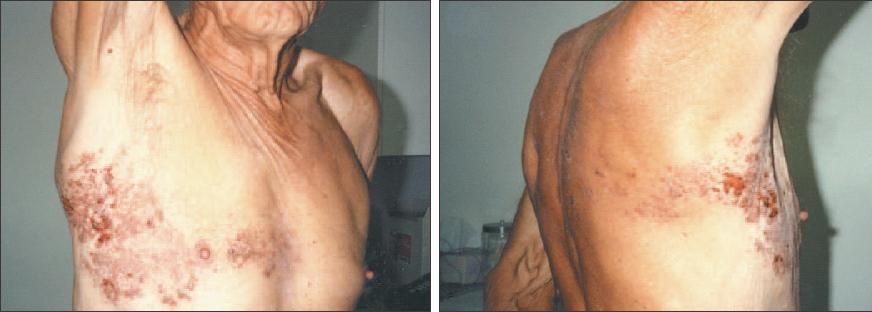
Images and case courtesy of William A. Hayes, MD.
Click here for the next image
Rarely does herpes zoster involve 2 dermatomes, as depicted here: the left T4 and T10. The painful, vesicular lesions on erythematous bases simultaneously began to develop along both dermatomes.

Image and case courtesy of Edwin J.Masters, MD, and J. Patrick Downey, MD.
Click here for the next image
A zoster eruption can present as a plaque of crusted pustules in a dermatome (top) or as urticaria-like plaques (bottom).
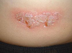
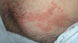
Images courtesy of Noah Scheinfeld, MD, JD.
Click here for the next image
The erosions on an erythematous base seen here are one of several typical manifestations of herpes zoster.
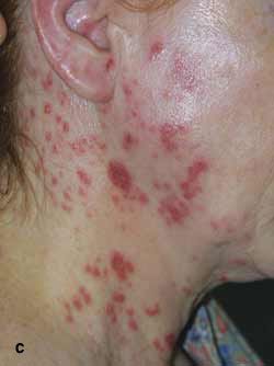
Image courtesy of Noah Scheinfeld, MD, JD.
Click here for the next image
These lesions on the distal thigh of a middle-aged woman featured erythema with irregular faded borders and central redness (top) with very fine papules (bottom) over the L3 dermatome. Microscopically, lesions of herpes simplex, herpes zoster, and varicella are nearly identical. The degree of leukocytoclasia and hemorrhage seen in the specimen in this case strongly suggested herpes zoster and, to a lesser degree, varicella.


Case and images courtesy of Robert P. Blereau, MD.
Click here for the next image
These crusted lesions and skin erythema follow the distribution of the ophthalmic, or first division of the trigeminal (fifth cranial), nerve and are manifestations of acute herpes zoster ophthalmicus. The ophthalmic infection accounts for 10% to 25% of all cases of herpes zoster.
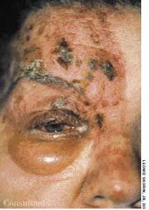
Photo courtesy of Leonid Skorin, Jr, MD.
Click here for the next image
This illustration of herpes zoster ophthalmicus shows the telltale involvement of the nasociliary nerve (Hutchinson sign).
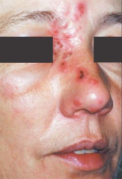
Photo courtesy of Sunita Puri, MD.
Click here for the next image
The distribution of these crusted lesions corresponds to the ophthalmic branch of the trigeminal nerve. Examination of the right eye revealed an edematous cornea with dendrites and anterior chamber inflammation, all findings consistent with ophthalmic zoster dermatitis.
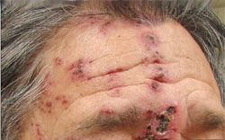
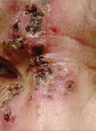
Photos and case courtesy of Khiem T. Tran, MD,
Andrew S. Qualm, OD, and Marcia A. Shannon, FNP.
Click here for the next image
Redness and swelling of the left cheek, chin, and ear were initially treated as cellulitis in this 51-year-old man. The outbreak along the maxillary and mandibular branches of the left trigeminal nerve, however, raised the suspicion of a varicella-zoster virus infection.
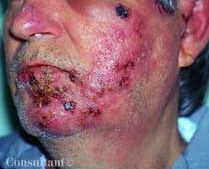
Photo and case courtesy of Robert P. Blereau, MD.
Click here for the next image
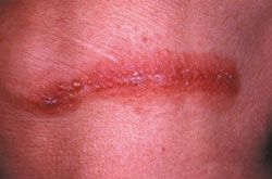
Examination of this acute painful rash on the chest wall of an 8-year-old girl revealed grouped vesicles with an erythematous base in a linear distribution along the T5 dermatome. The child was not vaccinated with varicella and had had chickenpox 3 years earlier. Herpes zoster is not common in children, but it can occur.
Click here to return to the first image.
