- Clinical Technology
- Adult Immunization
- Hepatology
- Pediatric Immunization
- Screening
- Psychiatry
- Allergy
- Women's Health
- Cardiology
- Pediatrics
- Dermatology
- Endocrinology
- Pain Management
- Gastroenterology
- Infectious Disease
- Obesity Medicine
- Rheumatology
- Nephrology
- Neurology
- Pulmonology
Foreign-Body Aspiration in a Middle-Aged Woman

A 52-year-old woman presented with a 1-week history of fever; a dry, irritating cough; and chest pain. She had nonpleuritic pain in the lower left side of the chest. There was no history of hemoptysis.
Findings from the general physical examination were normal. Examination of the respiratory system revealed impaired air entry at the base of the left lung; no other sounds were heard.
Results of routine laboratory tests, including hemogram and biochemistry, were normal. A chest radiograph showed a left-sided pleural effusion. The patient underwent decortication for left-sided pleural empyema. She remained on mechanical ventilation immediately following the surgery for respiratory failure and subsequently underwent a tracheostomy when an attempt to wean her from the ventilator failed. After the tracheostomy, there was a collapse of the left lower lobe, which led to pneumonia. A CT scan of the chest without contrast revealed a radiopaque density in the left lower lobe bronchus. On the basis of radiologic findings, a diagnosis of left lower lobe foreign-body aspiration (FBA) was entertained.

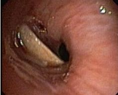
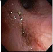
The patient underwent flexible bronchoscopy, which revealed a foreign body in the intermediate bronchus (Figure 1). The foreign body, which was found to be a piece of bone that the patient had aspirated while eating, was removed through the tracheostomy site with the bronchoscope. The bronchus area appeared edematous and erythematous (Figure 2). She was slowly weaned off the ventilator as her lung condition improved.
Discussion
FBA can lead to significant morbidity. In the United States, FBA is responsible for approximately 500 to 2000 deaths per year, half to three-quarters of which are in children younger than 4 years. Children younger than 4 years and adults older than 50 years account for most cases of FBA.1
In children, aspiration of foreign bodies is most common between the ages of 3 months and 6 years because of their curiosity, tendency to put objects in their mouth, and lack of molar teeth. Fifty percent of cases of FBA occur in children younger than 2 years, and 90% of cases occur in children younger than 4 years.1 Balloons, balls, and small toy parts are the most commonly aspirated items. Pieces of food, particularly hot dogs and peanuts, are frequently aspirated by toddlers.
FBA in adults with a normal swallowing reflex is rare. Most cases in adults are seen in the sixth or seventh decade of life, when airway protective mechanisms may function inadequately. Risk factors that may lead to aspiration include neurologic dysfunction, trauma with loss of consciousness, facial trauma, intubation, dental procedures, underlying pulmonary disease, impaired consciousness, alcohol consumption, and sedative use. Sources of the foreign bodies in adults include bone fragments, vegetable matter, broncholiths, part of a dental prosthesis, and tablets.
Upper airway involvement may cause complete obstruction at the trachea or larynx, or it may only cause an abnormality of the voice. Complete obstruction of a bronchus will cause atelectasis, while partial obstruction may cause emphysema, pneumothorax, or pneumomediastinum. Aspirated vegetable matter may produce a chronic suppurative pneumonitis. In many cases, the aspiration event is followed by cough and wheezing that may persist, usually without respiratory distress. Our case was unusual because the foreign body resulted in empyema and, subsequently, collapse of the left lower lobe, which led to development of postobstructive pneumonia.
In an analysis of 43 consecutive adult patients with FBA,2 chronic cough was present in 67%, hemoptysis in 23%, fever in 19%, and dyspnea in 16%, whereas only 7% had a history of choking.2 As seen in our case, a history of FBA is not always present. Therefore, the possibility of an inhaled foreign body should always be considered when there is sudden onset of choking and intractable cough with or without vomiting.
MAKING THE DIAGNOSIS
Clinical features
The diagnosis of FBA can be difficult, especially if the patient does not recall an aspiration episode. FBA has variable clinical manifestations, ranging from trivial symptoms to irreversible lung damage and life-threatening infection, atelectasis, and massive hemoptysis. Patients may present with a history of fever, breathlessness, or wheezing or with features of a nonresolving pneumonia. On physical examination, these patients may have decreased breath sounds on the side with the foreign body or localized wheezing, or they may be asymptomatic. The clinical triad that is considered to be diagnostic of FBA consists of wheezing, coughing, and diminished or absent breath sounds.
Imaging
The radiologic evaluation should start with anteroposterior and lateral views of the chest and neck. Although plain films may be interpreted as normal, radiopaque foreign bodies may be present. Other, indirect signs of an airway foreign body include resorption atelectasis beyond the site of bronchial obstruction and the presence of pulmonary infiltrates, indicating an inflammatory reaction.
A CT scan is helpful in visualizing radiopaque foreign bodies and alveolar collapse. It can also demonstrate airway foreign bodies that are radiolucent on plain radiographs. CT scans can depict a foreign body within the lumen of the tracheobronchial tree and the 3-dimensional position of the foreign body within the thorax.3 The other major radiologic features of FBA are lobar atelectasis and lobar pneumonia.
Most foreign bodies lodge in the right main bronchus, intermediate bronchus, or right lower lobe bronchus. In the upright person, these locations are direct entry from the trachea. The left main bronchus is smaller than the right main bronchus and is slightly angled. In the supine person, material usually enters the right main bronchus but then falls into the orifice of the right upper lobe, which is dependent in the supine position. Less common locations are the left lower lobe bronchus and left main bronchus. Occasionally, the trachea and, rarely, the left lower segmental bronchi are the sites of obstruction.
When diagnosis is delayed, the potential complications of a retained foreign body include unresolving pneumonia, lung abscess, recurrent hemoptysis, and bronchiectasis. A chronic inflammatory reaction may develop, with granulation tissue formation around the inhaled foreign body that may mimic bronchogenic carcinoma.4
TREATMENT
Once an aspirated foreign body is detected on radiologic studies, or if the clinical suspicion is high, the patient should undergo bronchoscopy to confirm the diagnosis and to remove the foreign body. If the object can be removed during bronchoscopy, further invasive procedures may be avoided. Rigid bronchoscopy, laryngoscopy,5 flexible bronchoscopy, and thoracotomy are the methods typically used to extract a foreign body from the tracheobronchial tree.
In children, rigid bronchoscopy using a ventilation bronchoscope offers good visualization and is the preferred mode for the removal of a tracheobronchial foreign body.1 The rigid instrument affords a better view of the foreign body and allows the use of a much wider range and size of forceps for removal than does the flexible fiberoptic bronchoscope.
In adults, a flexible fiberoptic bronchoscope can be used successfully to remove most foreign bodies.6 This type of bronchoscopy is easy to perform and can be done under local anesthesia; thus, it is preferred by most bronchoscopists.7 This procedure is also useful when the foreign body is more peripherally located.8 The most useful instruments for removal are alligator forceps and the wire basket.9 Fluoroscopy can help locate distal foreign bodies in the lung.10
Occasionally, an endobronchial foreign body extraction is accomplished with the use of a Fogarty balloon catheter. The foreign body is first located and visualized by a flexible fiberoptic bronchoscope. The Fogarty catheter is then deployed through the instrument channel of the bronchoscope beyond the foreign body. At this point, the balloon tip is inflated and pulled proximally against the object until it is wedged against the bronchoscope. The bronchoscope, foreign body, and Fogarty catheter are then withdrawn as a unit.11
Despite the advances in optical technology, proper training and experience are crucial to optimize the outcome and minimize the risk of complications in tracheobronchial foreign-body removal.
Flexible bronchoscopy can also be used in patients who are already intubated at the time of initial presentation. If bronchoscopy is performed for removal of a foreign body, positive pressure ventilation should be avoided because it may result in more distal impaction of the foreign body and may lead to an exacerbation of any air trapping distal to the foreign body.
The rigid bronchoscope remains the ideal instrument for retrieval of large foreign bodies from the main bronchi because it ensures better access, oxygen delivery, and easy passage of grasping forceps. In long-neglected cases in which a foreign body cannot be removed with the bronchoscope, a surgical approach may be required.
At the time of bronchoscopy, following removal of the foreign body, a “second look” should always be undertaken to identify any additional foreign bodies, complications of FBA, and iatrogenic injuries. Postoperatively, most patients should be observed overnight, if possible, for airway distress. Antibiotics should be considered in patients with fever or preoperative pneumonia and in those whose foreign body was vegetable matter. In cases in which the foreign body has been present for long time and there is fibrosis or impaction and removal with a bronchoscope is not possible, and a thoracotomy may be required.1
COMPLICATIONS
Complications of FBA or the surgical treatment of FBA include airway edema, respiratory distress, postobstructive pneumonia, postobstructive hypoxemia, hemoptysis, airway perforation, airway stenosis, development of an inflammatory polyp at the site of lodgment, or scarring. Bronchiectasis and mucoid impaction may result from prolonged retention of a bronchial foreign body.
References:
References
1. Baharloo F, Veyckemans F, Francis C, et al. Tracheobronchial foreign bodies: presentation and management in children and adults. Chest. 1999;115:1357-1362.
2. Chen CH, Lai CL, Tsai TT, et al. Foreign body aspiration into the lower airway in Chinese adults. Chest. 1997;112:129-133.
3. Zissin R, Shapiro-Feinberg M, Rozenman J, et al. CT findings of the chest in adults with aspirated foreign bodies. Eur Radiol. 2001;11:606-611.
4. Nigam BK. Bronchial foreign body masquerading as a lung carcinoma. Indian J Chest Dis Allied Sci. 1990;32:43-47.
5. Umapathy N, Panesar J, Whitehead BF, Taylor JF. Removal of a foreign body from the bronchial tree-a new method. J Laryngol Otol. 1999;113:851-853.
6. Cunanan OS. The flexible fibreoptic bronchoscope in foreign body removal. Experience in 300 cases. Chest. 1978;73(5 suppl):725-726.
7. Limper AH, Prakash UB. Tracheobronchial foreign bodies in adults. Ann Intern Med. 1990;112:604-609.
8. Zavala DC, Rhodes ML. Foreign body removal: a new role for the fiberoptic bronchoscope. Ann Otol Rhinol Laryngol. 1975;84(5, pt 1):650-656.
9. Debeljak A, Sorli J, Music E, Kecelj P. Bronchoscopic removal of foreign bodies in adults: experience with 62 patients from 1974-1998. Eur Respir J. 1999;14:792-795.
10. Nalaboff KM, Solis JL, Simon D. Endobronchial foreign body extraction: a new interventional approach. Chest. 2001;120:1402-1405.
11. Kosloske AM. The Fogarty balloon technique for the removal of foreign bodies from the tracheobronchial tree. Surg Gynecol Obstet. 1982;155:72-73.
Â
