- Clinical Technology
- Adult Immunization
- Hepatology
- Pediatric Immunization
- Screening
- Psychiatry
- Allergy
- Women's Health
- Cardiology
- Pediatrics
- Dermatology
- Endocrinology
- Pain Management
- Gastroenterology
- Infectious Disease
- Obesity Medicine
- Rheumatology
- Nephrology
- Neurology
- Pulmonology
Fatal HIV-Associated Anaplastic Large-Cell Lymphoma
Lymphoma is a well-known complication of HIV infection. Such AIDS-defining lymphomas are usually aggressive B-cell lymphomas. However, epidemiological data have also linked HIV infection with an increased risk of T-cell lymphoma.
Lymphoma is a well-known complication of HIV infection. Such AIDS-defining lymphomas are usually aggressive B-cell lymphomas. However, epidemiological data have also linked HIV infection with an increased risk of T-cell lymphoma.1 Therefore, it is easy to appreciate why the number of AIDS-related T-cell lymphoma case reports has increased in the literature.2 We present an interesting case of fatal anaplastic large-cell lymphoma of T-cell phenotype associated with Epstein-Barr virus (EBV) infection in a patient with AIDS.
A 42-year-old Hispanic man with HIV infection diagnosed 4 years earlier had a recent medical history significant for Mycobacterium avium-intracellulare pneumonia. He had been treated with ethambutol and azithromycin and also had been given Pneumocystis jiroveci pneumonia prophylaxis. He had been nonadherent to his antiretroviral treatment regimen.
On presentation, he complained of vomiting, diarrhea, 1 week of fever (temperature of 38.8°C [101.9°F]), weakness, and a 2-day history of urinary incontinence unrelated to a neurological cause. His absolute CD4+ cell count was 63/µL, and his HIV RNA level was 227,637 copies/mL. Physical examination revealed dehydration, oral thrush, parotid enlargement, and nontender left cervical lymphadenopathy.
Results of laboratory testing showed a white blood cell count of 2300/µL, with 13% lymphocytes (normal, 15% to 43%); hemoglobin level of 8.8 g/dL (normal, 14 to 18); platelet count of 121,000/µL (normal, 150,000 to 460,000); abnormal findings on urinalysis (3+ albuminuria, 3+ hemoglobinuria, and casts); normal electrolyte levels and renal function; serum albumin level of 2.6 g/dL (normal, 3.4 to 4.8); total bilirubin level of 0.1 mg/dL (normal, 0.0 to 1.0); and lactate dehydrogenase level of 878 U/L (normal, 94 to 250). M avium-intracellulare was found in a bronchoalveolar lavage specimen. A blood culture grew Bacteroides thetaiotaomicron.
Despite cefepime therapy, the patient remained febrile. Respiratory distress developed, and he became hypotensive with ensuing acute renal and liver failure. Diffuse airspace disease was noted on a chest radiograph. He died soon afterward, and an autopsy was performed.
Postmortem examination revealed anaplastic large-cell lymphoma involving his cervical, mediastinal, periaortic, mesenteric, and peripancreatic lymph nodes as well as multiple organs, including the lungs, heart, kidneys, adrenal glands, esophagus, stomach, and liver (Figures 1, 2, 3, and 4). His cervical lymph nodes measured up to 2.5 cm. In addition, several deep visceral lymph nodes were enlarged and measured up to 1.0 cm; most of these lymph nodes were grossly replaced by lymphoma. His lungs were firm and noncrepitant on gross examination and were notable for multiple white tumor nodules bilaterally on the visceral pleura and within the parenchyma (Figure 1, left). Apart from extensive pulmonary involvement by lymphoma, there was also diffuse alveolar damage (Figure 1, right) but no evidence of mycobacterial infection.
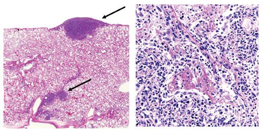
Figure 1. Left: Lymphoma nodules (arrows) are shown on the visceral pleura and in the lung parenchyma along the septum (hematoxylin and eosin stain). Right: Anaplastic lymphoma cells are seen extensively infiltrating the lung parenchyma admixed with intra-alveolar eosinophilic (pink) fibrin deposition in keeping with an exudative (acute) phase of diffuse alveolar damage (hematoxylin and eosin stain, original magnification ×400).
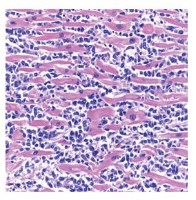
Figure 2. Anaplastic lymphoma cells can be seen diffusely infiltrating between myocytes in the heart (hematoxylin and eosin stain, original magnification ×400).
The lymphoma cells were immunoreactive for the T-cell markers CD45RO and CD30 (Figure 5) but failed to stain with CD20, CD79a, PAX5, CD3, CD15, CD56, CD57, ALK, epithelial membrane antigen, and latent nuclear antigen 1 (for human herpesvirus 8). In situ hybridization for EBV-encoded RNA was strongly positive. T-cell receptor (T-gamma) gene rearrangement by polymerase chain reaction analysis was positive for a clonal T-cell gene rearrangement.
The risk of T-cell lymphoma has been estimated to be 15-fold higher in HIV-positive persons. Although HIV-associated T-cell lymphoma was recognized relatively early in the AIDS epidemic,3-5 its occurrence in the pre-HAART era was believed to be rare.6 Many earlier cases of T-cell lymphoma have been grouped with B-cell lymphomas in larger series of AIDS-related lymphoma. The exact pathogenesis of T-cell lymphoma in the setting of HIV infection is uncertain but appears to be related to both immunosuppression and viral coinfection (eg, with EBV).2
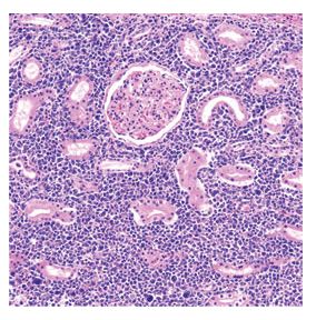
Figure 3. The kidney interstitium is densely packed with malignant lymphocytes growing between renal glomeruli and tubules (hematoxylin and eosin stain, original magnification ×200).
Several concomitant HIV-induced T-cell abnormalities are believed to predispose patients to natural killer/T-cell lymphomas. For example, some authors believe that the preponderance of CD8+ lymphocytes seen with chronic HIV infection may facilitate their neoplastic transformation.7 While the frequency of EBV in anaplastic large-cell lymphoma not associated with HIV is low,8 there have been several reports showing a high frequency of EBV detection in anaplastic large-cell lymphoma associated with HIV infection.9-11 It has been suggested by some authors that EBV infection may mediate progression of other non-Hodgkin lymphomas to CD30+ anaplastic large-cell lymphoma.12 Interestingly, cases of anaplastic large-cell lymphoma associated with EBV infection appear not to be associated with ALK gene expression because of the classic chromosomal translocation involving chromosomes 2 and 5, ie, t(2;5).13 In support of this observation, the EBV-associated anaplastic large-cell lymphoma in our HIV-positive patient was ALK-negative.
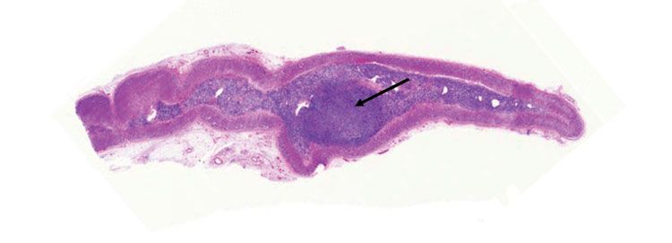
Figure 4. A large lymphoma nodule (arrow) is shown within the medulla of an adrenal gland (hematoxylin and eosin stain).
AIDS-related T-cell lymphomas usually present at an advanced lymphoma stage in patients with marked immunosuppression and prior AIDS-defining illness.2 Such patients commonly have B symptoms (eg, weight loss, night sweats, fever), lymphadenopathy, and extranodal disease. Many of the clinical manifestations of T-cell lymphoma, such as fever, can be related to cytokine expression from lymphoma cells. In general, systemic AIDS-related T-cell lymphomas exhibit an aggressive clinical course with a poor prognosis.
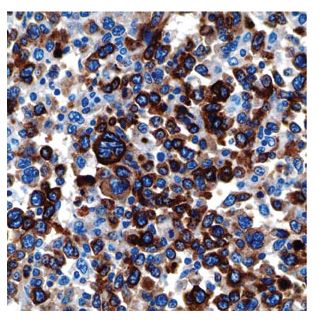
Figure 5. Lymph node effacement caused by anaplastic lymphoma cells (immunohistochemical stain for CD30, original magnification ×600).
In one series involving 4 patients with CD30+ T-cell anaplastic large-cell lymphoma, similar to that seen in our patient, investigators reported aggressive disease with patients dying within a median of 3 months from their lymphoma diagnosis.14 Factors that likely contribute to the aggressive clinical behavior of these malignancies include advanced clinical lymphoma stage at presentation, marked HIV-related immunosuppression, and chemotherapeutic resistance. A standard therapeutic approach is required in order to address these emerging AIDS-defining T-cell neoplasms, for which prognosis remains poor even in the HAART era.2
Finally, autopsy rates for patients dying in hospitals have declined dramatically over the past 50 years.15 This case clearly demonstrates the clinical value of postmortem examination. Autopsies should be reasserted as a tool for investigation and quality improvement in modern medicine.
References:
References1. Biggar RJ, Engels EA, Frisch M, Goedert JJ; AIDS Cancer Match Registry Study Group. Risk of T-cell lymphomas in persons with AIDS. J Acquir Immune Defic Syndr. 2001;26:371-376.
2. Castillo J, Pantanowitz L. HIV-associated NK/T-cell lymphomas: a review of 93 cases [abstract]. Blood. 2007;18:1013A. Abstract 3457.
3. Kobayashi M, Yoshimoto S, Fujishita M, et al. HTLV-positive T-cell lymphoma/leukaemia in an AIDS patient. Lancet. 1984;1:1361-1362.
4. Nasr SA, Brynes RK, Garrison CP, Chan WC. Peripheral T-cell lymphoma in a patient with acquired immune deficiency syndrome. Cancer. 1988;61:947-951.
5. Sternlieb J, Mintzer D, Kwa D, Gluckman S. Peripheral T-cell lymphoma in a patient with the acquired immunodeficiency syndrome. Am J Med. 1988;85:445.
6. Janier M, Katlama C, Flageul B, et al. The pseudo-Sézary syndrome with CD8 phenotype in a patient with the acquired immunodeficiency syndrome (AIDS). Ann Intern Med. 1989;110:738-740.
7. Ruco LP, Di Napoli A, Pilozzi E, et al. Peripheral T cell lymphoma with cytotoxic phenotype: an emerging disease in HIV-infected patients? AIDS Res Hum Retroviruses. 2004;20:129-133.
8. Herling M, Rassidakis GZ, Jones D, et al. Absence of Epstein-Barr virus in anaplastic large cell lymphoma: a study of 64 cases classified according to World Health Organization criteria. Hum Pathol. 2004;35:455-459.
9. Carbone A, Gloghini A, Volpe R, et al. High frequency of Epstein-Barr virus latent membrane protein-1 expression in acquired immunodeficiency syndrome-related Ki-1 (CD30)-positive anaplastic large-cell lymphomas. Italian Cooperative Group on AIDS and Tumors. Am J Clin Pathol. 1994;101:768-772.
10. Nosari A, Cantoni S, Oreste P, et al. Anaplastic large cell (CD30/Ki-1+) lymphoma in HIV+ patients: clinical and pathological findings in a group of ten patients. Br J Haematol. 1996;95:508-512.
11. Fätkenheuer G, Hell K, Roers A, et al. Spontaneous regression of HIV associated T-cell non-Hodgkin’s lymphoma with highly active antiretroviral therapy. Eur J Med Res. 2000;5:236-240.
12. DiGiuseppe JA, Wu TC, Zehnbauer BA, et al. Epstein-Bar virus and progression of non-Hodgkin’s lymphoma to Ki-1-positive, anaplastic large cell phenotype. Mod Pathol. 1995;8:553-559.
13. Agarwal S, Ramanathan U, Naresh KN. Epstein-Barr virus association and ALK gene expression in anaplastic large-cell lymphoma. Hum Pathol. 2002;33:146-152.
14. Chadburn A, Cesarman E, Jagirdar J, et al. CD30 (Ki-1) positive anaplastic large cell lymphomas in individuals infected with the human immunodeficiency virus. Cancer. 1993;72:3078-3090.
15. Hooper JE, Geller SA. Relevance of the autopsy as a medical tool: a large database of physician attitudes. Arch Pathol Lab Med. 2007;131:268-274.
Â
