- Clinical Technology
- Adult Immunization
- Hepatology
- Pediatric Immunization
- Screening
- Psychiatry
- Allergy
- Women's Health
- Cardiology
- Pediatrics
- Dermatology
- Endocrinology
- Pain Management
- Gastroenterology
- Infectious Disease
- Obesity Medicine
- Rheumatology
- Nephrology
- Neurology
- Pulmonology
Diabetes Disorders-A Photo Essay
More than 90% of patients with diabetes mellitus receive their care from primary care physicians. This compact slide show provides visual presentations of a range of DM-related problems.
Acute stage Charcot foot is seen in a 55-year-old man with a significant past medical history of type 2 diabetes mellitus. He shows peripheral neuroarthropathy with a 1-week history of a hot, swollen right foot. Charcot neuroarthropathy is a major cause of morbidity for patients with DM.

Image courtesy of Jackie Pham, PMS-IV, Bora Rhim, DPM, and Jonathan Labovitz, DPM.
Click here for the next image
This patient has a 2-year history of red plaque with a yellow atrophic center on the leg, which has ulcerated over the past 3 months. There is a strong family history of diabetes mellitus. The patient has necrobiosis lipoidica, a condition associated with a personal or family history of DM. The waxy, atrophic appearance of the lesion, with its yellow center, is diagnostic; ulceration is a late complication.
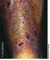
Image courtesy of Norman Levine, MD.
Click here for the next image
A 50-year-old African American woman with type 2 diabetes mellitus and hypertension was admitted with bilateral knee and thigh pain and swelling of both knees. MRI showed extensive edema in the distal thigh and gastrocnemius muscles and in subcutaneous fat. Fluid was seen at the short head of the left biceps femoris. The findings were consistent with diabetic myonecrosis, a rare complication of uncontrolled DM that should be suspected in patients with type 2 DM who present with thigh pain or knee effusions.

Images courtesy of Kiran Kondaveeti and Cuc Mai, MD.
Click here for the next image
In a patient with diabetes mellitus, a small, untreated blister can easily develop into a full-thickness ulceration. A diabetic ulcer, whether of neuropathic or ischemic origin (as seen here), could be limb-threatening-it can easily progress to sepsis or gangrene and eventuate in amputation. The main risk factors associated with ulceration and amputation are neuropathy, vascular disease, impaired immune response, and structural deformities.
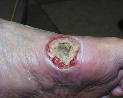
Image courtesy of Robert G. Frykberg, DPM, MPH and Donald Curtis, DPM.
Click here for the next image
Hypertension of a patient’s metatarsal phalangeal joint is seen in this clawed right hallux. Note the superficial ulcer (a result of irritation from shoe contact) over the hyperflexed interphalangeal joint. The extensor tendon “bowstrings” (arrow) over the MTPJ because of joint hyperextension. Clawed toes are common in patients with diabetes mellitus.

Image courtesy of Michael B. Strauss, MD and Stuart S. Miller, MD.
Click here for the next image
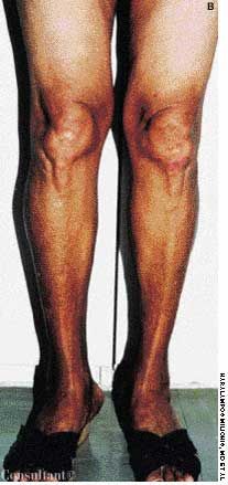
Diabetes mellitus had been diagnosed in this 58-year-old woman, who claimed that her skin had darkened significantly over the previous 5 years. Hepatomegaly was noted. Histopathological examination of a liver biopsy specimen was consistent with hemochromatosis, or bronze diabetes, a genetic disorder caused by an overstorage of iron in the body that leads to organ damage.

Images courtesy of Haralampos Milionis, MD, George Liamis, MD, and Moses Elisaf, MD.
Click here for the next image
Patients with diabetes mellitus, especially those whose feet show signs of dysvascularity, are particularly susceptible to fungal infections and dystrophic changes in the toenails. Note this patient’s long, fungus-thickened toenails and the mildly dry and scaly skin.

Image courtesy of Michael B. Strauss, MD and Stuart S. Miller, MD.
Click here for the next image
A 32-year-old woman with insulin-dependent diabetes mellitus noted a painful erosion at the site of “the rose tattoo,” which she had gotten 5 days earlier. A culture isolated Staphylococcus aureus, confirming the clinical impression of staphylococcal cellulitis. Patients with DM are predisposed to staphylococcal infection.
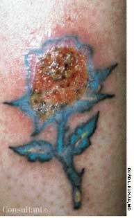
Image courtesy of David L. Kaplan, MD.
Click here to return to the first image.
