- Clinical Technology
- Adult Immunization
- Hepatology
- Pediatric Immunization
- Screening
- Psychiatry
- Allergy
- Women's Health
- Cardiology
- Pediatrics
- Dermatology
- Endocrinology
- Pain Management
- Gastroenterology
- Infectious Disease
- Obesity Medicine
- Rheumatology
- Nephrology
- Neurology
- Pulmonology
The Changing Face of Anal Cancer
Cancer of the anal canal is a relatively uncommon disease in the United States. It accounts for about 2% of the cancers of the GI tract; about 5000 cases will be diagnosed this year. Squamous cell carcinoma of the anus (anal SCC) is of particular interest to the infectious disease specialist because it is one of the cancers associated with HIV infection in men who have sex with men (MSM).
Cancer of the anal canal is a relatively uncommon disease in the United States. It accounts for about 2% of the cancers of the GI tract; about 5000 cases will be diagnosed this year. Squamous cell carcinoma of the anus (anal SCC) is of particular interest to the infectious disease specialist because it is one of the cancers associated with HIV infection in men who have sex with men (MSM). During the HAART era, the incidence of anal cancer in MSM has risen sharply, with about 70 new diagnoses per 100,000 patients.1 As a result, anal SCC is one of the most common solid-organ malignancies seen in male HIV-infected patients, and it should be a focus for any clinician taking care of this patient population.
Although HIV is not the primary etiological agent in anal SCC, infection with HIV is a marker for coinfection with other sexually transmitted diseases––including human papillomavirus (HPV) infection. HPV is the sexually transmitted DNA virus that causes anal SCC. HPV subtypes 16 and 18 have a higher malignant potential than other HPV subtypes. Similar to HPV-infected cervical cells, HPV-infected anal mucosal cells undergo a temporal progression from anal intraepithelial neoplasia (AIN) to invasive anal cancer. Interestingly, coinfection with HIV may actually augment the carcinogenicity of HPV by mechanisms other than immunosuppression. HIV produces the Tat protein, which in vitro up-regulates the transcription of the E6 and E7 proteins by HPV. These 2 proteins promote tumorigenesis by blocking cell cycle arrest and apoptosis in HPV-infected cells.2
Before the HIV epidemic, anal SCC occurred primarily in menopausal women. These patients typically complained of rectal bleeding or pain, and the primary finding on physical examination was an anal mass (Figure 1).3 In this presentation, the examining physician should not only document where in the anus the mass is located (right or left, anterior or posterior, above or below the dentate line) but also note the consistency of the tumor (eg, firm or soft) and whether it is fixed to deeper structures. The clinician should also inspect the inguinal folds for lymphadenopathy or other signs of metastatic disease.
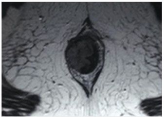
Figure 1. Axial MRI of pelvis showing anal mass. (Reproduced with permission from Clark MA et al. Lancet Oncol. 2004.3)
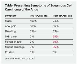
The clinical presentation of anal SCC has shifted in the past 2 decades as the number of affected persons has increased. Because of the increasing number of patients with HIV-associated anal cancers, anal cancer now not only develops at a younger age than it did in the pre-HAART era (mean age at diagnosis, 42 vs 58 years, respectively) but also can present in quite atypical ways. Figures 2, 3, and 4 show a variety of clinical presentations of anal SCC in HIV-positive patients from our recent series.4 Figure 2 shows a bleeding mass, Figure 3 depicts an ulcerated lesion, and Figure 4 shows an anal SCC presenting as a fistula-in-ano at the 3 o’clock position. Figure 4 also shows a protruding internal hemorrhoid at 6 o’clock and a plaque-like wart at 9 o’clock.
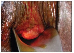
Figure 2.Intraoperative photo of a squamous cell carcinoma presenting as a bleeding anal mass.
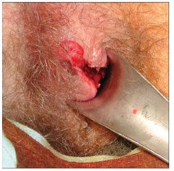
Figure 3.Intraoperative photo of anulcerated squamous cell carcinoma of the anus.
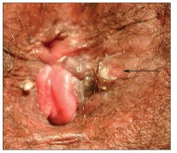
Figure 4. Intraoperative photo of a fistula-in-ano that was proved to be anal squamous cell carcinoma on biopsy.
The Table demonstrates that these atypical presentations of anal SCC are just as common as painful or bleeding anal masses in the HIV-infected population. Because of such findings, a clinician treating MSM must have a high index of suspicion in any HIV-positive person presenting with anorectal complaints. Definitive diagnosis can be made pathologically from a biopsy specimen obtained during rectal examination, either under local or general anesthesia. Imaging studies-such as pelvic or endorectal MRI-can determine tumor depth and regional lymph node status. Also, a torso CT scan is usually obtained to identify metastatic disease. An HIV test should be offered to patients in whom this infection has not previously been diagnosed to identify the subset who may benefit from antiretroviral therapy.
Patients with AIN or invasive anal SCC should be referred to a colorectal surgeon for definitive therapy. AIN and stage I anal SCC (ie, tumors 2 cm or less) can be treated with wide local excision alone, with reported 5-year survival rates of up to 100%.5
Traditionally, more advanced tumors required an abdominoperineal rectal resection with a permanent end colostomy: expected 5-year survival rates ranged between 50% and 70%. Since the landmark paper by Nigro and colleagues6 in 1974, however, the combination of chemotherapy (mitomycin C, fluorouracil) and local, low-dose radiation has supplanted surgical treatment. The Nigro protocol results in a cure rate from 80% to 90% and has replaced abdominoperineal rectal resection as the standard of care. Surgical treatment has been relegated to cases of persistent or recurrent disease, or when a patient has a contraindication to chemotherapy or radiation therapy.
Hypervigilant monitoring of high-risk persons is another form of cancer prevention and disease management. Several groups have investigated the benefit of routine Papanicolaou (Pap) tests in the HIV-positive MSM cohort, given the success of routine screening in reducing cervical cancer in women. Recently, Chiao and associates7 concluded that annual Pap screening in HIV-positive MSM was financially reasonable and that screening every 2 or 3 years in HIV-negative MSM was also cost-effective. On the other hand, Pap tests are not useful in patients who have already been treated for anal SCC, because changes in the mucosa from radiation produce numerous false-positive results.
No potential conflict of interest relevant to this article was reported by the authors.
References:
References1. Melbye M, Cote TR, Kessler L, et al. High incidence of anal cancer among AIDS patients. The AIDS/Cancer Working Group. Lancet. 1994;343:636-639.
2. Palefsky JM. Anal squamous intraepithelial lesions: relation to HIV and human papillomavirus infection. J Acquir Immune Defic Syndr. 1999;21(suppl 1):S42-S48.
3. Clark MA, Hartley A, Geh JI. Cancer of the anal canal. Lancet Oncol. 2004;5:149-157.
4. Kundu R, Nagle D, Henry D, et al. Squamous cell carcinoma of anal canal: a changing entity in the era of HIV disease. Annual Meeting of the American Society of Colon and Rectal Surgeons; June 3-7, 2006; Seattle.
5. Welton ML, Varma MG. Anal cancer. In: Wolff BG, Fleshman JW, Beck DE, et al, eds. The ASCRS Textbook of Colon and Rectal Surgery. New York: Springer; 2007:482-500.
6. Nigro ND, Vaitkevicius VK, Considine B Jr. Combined therapy for cancer of the anal canal: a preliminary report. Dis Colon Rectum. 1974;173:354-356.
7. Chiao EY, Giordano TP, Palefsky JM, et al. Screening HIV-infected individuals for anal cancer precursor lesions: a systematic review. Clin Infect Dis. 2006;43:223-233.
