- Clinical Technology
- Adult Immunization
- Hepatology
- Pediatric Immunization
- Screening
- Psychiatry
- Allergy
- Women's Health
- Cardiology
- Pediatrics
- Dermatology
- Endocrinology
- Pain Management
- Gastroenterology
- Infectious Disease
- Obesity Medicine
- Rheumatology
- Nephrology
- Neurology
- Pulmonology
Ascites as the Presenting Sign of Hypereosinophilic Syndrome
Eosinophilic ascites is an extremely rare presenting sign of hypereosinophilic syndrome.
A 45-year-old man presented to a primary health clinic with a 3-week history of increasing abdominal girth and diffuse abdominal pain. He had gained 5 pounds within a week despite diminished appetite. He had a 2-week history of fevers (temperature as high as 38.6°C [101.5°F]) with night sweats.
His past medical history was notable for an ICU admission 9 months earlier for an unusual anaphylactic reaction that was attributed to sinusitis and a retropharyngeal mass. That admission had been complicated by acute renal failure and development of pulmonary emboli. CT scan findings at that time suggested mediastinitis, but an infectious disease workup was negative for HIV infection, hepatitis, tuberculosis, and systemic actinomycosis. A biopsy specimen of the retropharyngeal mass was positive for eosinophilic granuloma.

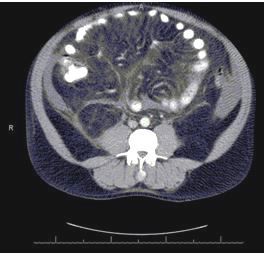
The patient was admitted for workup of abdominal pain and evaluation of ascites and bilateral pleural effusions noted on CT scan (Figure 1, left). There were no radiological findings of ischemic colitis, portal hypertension, hepatic vein thrombosis, or mesenteric ischemia. A complete blood cell count initially showed a hemoglobin level of 10.8 g/dL (normal range, 14-18 g/dL), a platelet count of 593,000/µL (normal range, 150,000-400,000/µL), and a normal white blood cell count of 91,000/µL, with eosinophilia (2300 cells/µL; normal <350 cells/µL); during the hospital course, the eosinophil count was as high as 4500/µL. Results of an iron panel were consistent with anemia of chronic disease. A comprehensive metabolic panel revealed BUN of 23 mg/dL (normal range, 8-23 mg/dL) and creatinine of 1.2 mg/dL (normal range, 0.6-1.2 mg/dL), with normal liver function. Erythrocyte sedimentation rate was elevated at 69 mm/h (normal range, 0-20 mm/h).

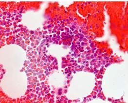
Paracentesis revealed ascitic fluid with negative Gram stain/culture, but with a serum ascites albumin gradient of 3.0 and absolute eosinophil count of 6200/µL. Bone marrow biopsy of the left iliac crest showed moderately hypercellular myeloid-predominant bone marrow with marked eosinophilia (Figure 2, left). Flow cytometry and chromosome analysis studies, specifically CD3, CD4, CD8, and CD34 expressions, were negative. Tests for stool ova and parasites and for serum Strongyloides antibodies were negative. Results of autoimmune serologic studies, including ANA, ANCA, and lupus anticoagulant, were negative. Results of quantitative immunoglobulin tests were unremarkable.
Hypereosinophilic syndrome was the diagnosis. A prednisone taper regimen was initiated. The abdominal pain resolved, and the patient was discharged 7 days later in stable condition. A month later, a punch biopsy was performed of a lesion on the patient’s thumb. Histology showed a mixed dermal inflammatory cell infiltrate, similar to that found in the biopsy of the retropharyngeal mass 10 months earlier. After adverse effects from the prednisone, hydroxyurea was substituted; the patient is now slowly being weaned off therapy.
Discussion
Hypereosinophilic syndrome has been defined as a myeloproliferative disorder that is1:
1. Characterized by persistent eosinophilia
2. Unexplained by a secondary cause, and
3. Associated with end-organ damage
It has been shown to affect various organ systems, most commonly hematologic, cardiovascular, and dermatologic, but also pulmonary, neurologic, and GI. A few cases have involved GI manifestations, including ascites, diarrhea, gastritis, colitis, and pancreatitis.2
Hypereosinophilic syndrome should not be confused with eosinophil-associated GI disorders. The latter is an umbrella term for organ-specific disorders; the former typically involves more than one organ. Nor should it be confused with GI eosinophilia secondary to other causes, such as parasitic infections, drug reaction, Churg-Strauss syndrome, systemic lupus erythematosus, and malignancy.3
We present a rare case in which the patient’s presenting sign of hypereosinophilic syndrome was ascites. Several of the documented cases of ascites have been secondary to liver disease, specifically Budd-Chiari syndrome and hepatic veno-occlusive disease.4,5 There are very few published cases in which eosinophilic ascites was considered the presenting symptom.6
First-line treatments are glucocorticoids with or without the tyrosine kinase inhibitor, imatinib,7 and if needed, the addition of a steroid-sparing drug such as hydroxyurea. The ultimate goal is to prevent progression to end-organ dysfunction by reducing eosinophilia and controlling symptoms and signs of the disease.
Take-Home Points
• Consider the diagnosis of hypereosinophilic syndrome in patients who present with ascites and paracentesis characterized by eosinophilia
• Patients with a primary eosinophilia-associated GI disorder who develop extra-GI symptoms should be evaluated for hypereosinophilic syndrome, because prognoses and therapies differ
• Patients with GI eosinophilia may have secondary causes that need to be ruled out before establishing a diagnosis of hypereosinophilic syndrome
• The goal of therapy in hypereosinophilic syndrome is to prevent progression of end-organ dysfunction
References
1. Chusid MJ, Dale DC, West BC, et al. The hypereosinophilic syndrome: analysis of fourteen cases with review of the literature. Medicine (Baltimore). 1975;54:1-27.
2. Gotlib J, Cools J, Malone JM 3rd, et al. The FIP1L1-PDGFRalpha fusion tyrosine kinase in hypereosinophilic syndrome and chronic eosinophilic leukemia: implications for diagnosis, classification, and management. Blood. 2004;103:2879-2891.
3. Simon D, Wardlaw A, Rothenberg ME. Organ-specific eosinophilic disorders of the skin, lung and gastrointestinal tract. J Allergy Clin Immunol. 2010;126:3-13.
4. Inoue A, Michitaka K, Shigematsu S, et al. Budd-Chiari syndrome associated with hypereosinophilic syndrome; a case report. Intern Med. 2007;46:1095-1100.
5. Kojima K, Sasaki T. Veno-occlusive disease in hypereosinophilic syndrome. Intern Med. 1995;34:1194-1197.
6. de Carpi JM, Rives S, Prada F, et al. Eosinophilic ascites as the first sign of idiopathic hypereosinophilic syndrome in childhood. J Clin Gastroenterol. 2007;41:864-865.
7. Gleich GJ, Leiferman KM, Pardanani A, et al. Treatment of hypereosinophilic syndrome with imatinib mesilate. Lancet. 2002;359:1577-1578.
