- Clinical Technology
- Adult Immunization
- Hepatology
- Pediatric Immunization
- Screening
- Psychiatry
- Allergy
- Women's Health
- Cardiology
- Pediatrics
- Dermatology
- Endocrinology
- Pain Management
- Gastroenterology
- Infectious Disease
- Obesity Medicine
- Rheumatology
- Nephrology
- Neurology
- Pulmonology
Alopecia, Ulcerations, and Ecchymotic Lesions: Take this Image IQ Test
A 22-year-old Filipino man with fever, lethargy, weakness, and malaise of 5 days' duration was brought to the emergency department by his family. Two days earlier, oral penicillin had been prescribed for streptococcal pharyngitis. The patient was unable to walk because of profound weakness. Circular and linear ecchymotic lesions were noted on his back.

1. Fever, weakness, and unusual ecchymotic lesions
A 22-year-old Filipino man with fever, lethargy, weakness, and malaise of5 days' duration was brought to the emergency department by his family. Twodays earlier, oral penicillin had been prescribed for streptococcal pharyngitis.The patient was unable to walk because of profound weakness. Circular andlinear ecchymotic lesions were noted on his back.
What could account for these lesions? Are they related to the patient'sother symptoms?
(Answer on next page.)

2. Small pink lesion on the nose
A 35-year-old man feared that thesmall pink lesion that spontaneouslyappeared on the edge of one nostrilwas cancer. The polypoid, superficial,ulcerated nodule first appeared 3 weeksearlier.
Does this look like a malignantlesion?
(Answer on next page.)

1. Fever, weakness, and unusual ecchymotic lesions: Ahead CT scan demonstrated a posterior fossa abscess andcerebral edema, which were confirmed by an MRI scan.On hospital day 2, tense swelling of the left thigh wasnoted; the total creatine kinase level was elevated. Culturesof blood and muscle biopsy specimens grew Streptococcuspyogenes; streptococcal myositis was diagnosed as well.
A suboccipital craniotomy to drain the cerebellar abscesswas performed, and a 6-week course of intravenousantibiotics was initiated. After 29 days in the hospital, thepatient was discharged; outpatient rehabilitation wasbegun for his ataxia and upper extremity weakness.
The lesions on the patient's back were unrelated tothe infectious process and its treatment. They are the resultof cupping and coining, folk remedies that were performedby his family. Cupping has been practiced bymany cultures, including the Greeks, Chinese, Vietnamese,and Native Americans.1-3 As recently as the 1920s, thepractice was still widespread in the United States and wasoften prescribed by physicians for a wide range of maladies,such as headache, GI disorders, menstrual disturbances,myopathies, arthropod bites, neuropathies, andbronchopulmonary and cardiac disease.2,3
In dry cupping, a heated container or cup is placedover the skin of the back, neck, buttocks, or chest to form avacuum, which results in hyperemia and ecchymosis. Asimilar procedure is used in wet cupping, except a break inthe skin is made before the container is placed; this allowsfor the vacuum to extract blood. Adherents believe that cuppingwithdraws diseased "humors" from the skin and causes"counter-irritation" that shunts blood from the engorgedviscus to the hyperemic skin.1-4
Coining
, a Vietnamese practice also known as cao gio,often is used to relieve fever, chills, and headache.
5
The edgeof a coin is rubbed along the ribs or spine to create areas ofecchymosis and hyperemia. The practice, which may be performedseveral times during an illness, is intended to "rubout the wind" or release excessive "air" that is believed to bethe cause of illness.
6
Coining also is thought to restore balanceof the "hot and cold" or "yin-yang" forces of the body.These lesions have been mistaken for child abuse, and legalaction has been taken against some parents.
7,8
Complications of these practices include hyperpigmentation,scarring, burns that are often secondary tospills of the flammable material on the patient, and superinfection.
3,9
A case of a 25-year-old woman with intracerebellarhemorrhage secondary to presumed hypersecretion ofcatecholamines and resultant malignant hypertension alsohas been reported. Here, the patient became drowsy andunresponsive while coining was being performed to treather headache. A head CT scan revealed a large cerebellarhematoma and acute obstructive hydrocephalus. The painassociated with coining may have prompted a catecholaminesurge that caused malignant hypertension and acerebellar hemorrhage.
5
It is conceivable that the primaryevent was an intracranial hemorrhage that caused theheadache, which was treated with coining.It is important to recognize the skin lesions associatedwith traditional and folk remedies and to be aware thatthese therapies may be practiced in lieu of-or in additionto-standard allopathic care. Fifty-eight percent of emigrantsfrom Southeast Asia use traditional health practices;about three quarters of these persons report relief ofsymptoms.
6
REFERENCES:1. King DF, Davis MW. Cupping: an erstwhile common modality of therapy. J AmAcad Dermatol. 1983;8:563.
2. Haller JS Jr. The glass leech. Wet and dry cupping practices in the nineteenthcentury. N Y State J Med. 1973;73:583-592.
3. Stoeckle DB, Carter RD. Cupping in New York State-1978: historic review.N Y State J Med. 1980;80:117-120.
4. Powers RD. Photo case: cupping lesions. Acad Emerg Med. 1997;4:160.
5. Ponder A, Lehman LB. "Coining" and "coning": an unusual complication ofunconventional medicine. Neurology. 1994;44:774-775.
6. Buchwald D, Panwala S, Hooton TM. Use of traditional health practices bySoutheast Asian refugees in a primary care clinic. West J Med. 1992;156:507-511.
7. Asnes RS, Wisotsky DH. Cupping lesions simulating child abuse. J Pediatr. 1981;99:267-268.
8. Sandler AP, Haynes V. Nonaccidental trauma and medical folk belief: a case ofcupping. Pediatrics. 1978;61:921-922.
9. Sagi A, Ben-Meir P, Bibi C. Burn hazard from cupping-an ancient universalmedication still in practice. Burns Incl Therm Inj. 1988;14:323-325.
(Case and photograph courtesy of Drs Timothy R. Hurtado and David A.Della-Giustina.)

2. Small pink lesion on the nose: Ashave biopsy was performed and confirmedthe suspected diagnosis of pyogenicgranuloma, a benign vascular lesionthat frequently occurs in childrenand young adults. The epidermal collarettearound the papule's base, thecentral ulceration, and the fairly rapiddevelopment suggested the diagnosis.Pyogenic granuloma is thought to resultfrom trauma.
These lobular capillary hemangiomasgrow rapidly; they are usuallyless than 1 cm in diameter. Most commonly,they arise on the head, neck,or extremities. Spontaneous regressioncan occur within 6 months. Surgicalexcision or electrodesiccation andcurettage may be performed; removal must be complete to avoid regrowth andrecurrence.
(Case and photograph courtesy of Dr Robert P. Blereau.)

3. Unusual pigmentation pattern
Asymptomatic, dark brown patchessuddenly appeared on the leg of a53-year-old man who was vacationing ata Caribbean resort. He had spent mostof his time sunbathing on the beach. Onreturning to the United States, he immediatelysought medical attention.
What is the likely cause of thehyperpigmentation?
(Answer on next page.)

4. Chest pain and blistered thumb
A 41-year-old man was brought to the emergency department with leftsidedchest pain of 2 hours' duration. Twelve months earlier, he had undergonecoronary artery bypass grafting (CABG). A long-term cocaine, ethanol, and tobaccouser, the patient had recently been released from a 90-day mandatory stayin a drug rehabilitation program. He was taking no medications.
Blood pressure was 160/100 mm Hg; heart rate, 90 beats per minute; respirationrate, 18 breaths per minute; oxygen saturation, 99%; and temperature,37.2C (99F). The ECG showed left ventricular hypertrophy, left atrial enlargement,and nonspecific ST-T wave changes but no acute ischemia. The troponin-I level was less than 0.3 µg/L; other laboratory values were normal. The urinedrug screen was positive for cocaine metabolites.
In addition to a prominent CABG scar and bilateral scars on his legs fromvein grafts, blisters were noted on the patient's right thumb.What do you suspect is responsible for these blisters?
(Answer on next page.)

3. Unusual pigmentation pattern: Further questioning revealed that the patienthad spilled lemon-flavored rum punch on his leg while he was sunbathing.The hyperpigmented path of the spilled drink can be traced down the patient'sleg below the hemline of the shorts.
This exchange supports the diagnosis of phytophotodermatitis, which resultsfrom exposure to sunlight after contact with photosensitizing plant psoralens (furocoumarins).Lemon, lime, parsley, celery, parsnip, and fig are among the fruits andvegetables that cause this reaction.
The hyperpigmentation usually fades over time; azelaic acid or tretinoinmay be tried.
(Case and photograph courtesy of Drs Athena Kaporis, Judy R. Anderson, and J. Elliot Paulson.)

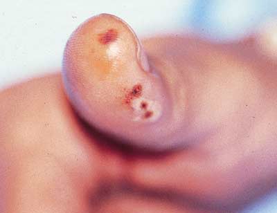
4. Chest pain and blistered thumb: Initially, the patientdenied recent cocaine use. When asked if his thumb injurywas caused by a lighter used for crack smoking, he admittedto having smoked the drug since leaving the rehabilitationprogram 9 days earlier. A disposable lighter used to heata crack pipe often causes either a callus or blister on theuser's thumb. This finding of "crack thumb" can be a valuableaid in detecting crack cocaine use.
After being given intravenous lorazepam, the patient'sblood pressure decreased to 143/89 mm Hg and his heartrate dropped to 74 beats per minute. The chest pain abated;he was discharged from the emergency departmentafter 6 hours.
(Case and photograph courtesy of Drs Kari Blaho and Stephen Winbery.)

5. Loss of leg hair
A hirsute 48-year-old man complained of the significant loss of leg hair duringthe past 6 months. He reported no other symptoms and continued to maintaina very active lifestyle. There was no weight loss, change in bowel habits, muscleweakening, or cold intolerance. Results of the physical examination were normal.
What do you infer about the cause of the hair loss?
(Answer on next page.)

6. An enlarging, painful ulceration on the temple
A 58-year-old man sought evaluation of an enlarging, painful ulceration onthe left temple that first arose 2 weeks earlier as a small papule. Recently, the patienthad been healthy; however, his history included unstable angina, a primaryseizure disorder, and narrow-angle glaucoma. His medications were diltiazem,phenytoin, aspirin, and pilocarpine. The patient smoked marijuana occasionally;he denied injection drug use.
What clues might reveal the diagnosis?
(Answer on next page.)

5. Loss of leg hair: Laboratory tests yielded elucidatingdata: the patient's thyroid-stimulating hormone level was222 µIU/mL and his creatine kinase level was 1500 U/L.Hypothyroidism was diagnosed. The patient's conditionresponded well to replacement therapy.
(Case and photograph courtesy of Dr Rebecca Galante.)

6. An enlarging, painful ulceration on the temple: The1.5-cm, firm, raised mass with a 3-mm central ulceration featuredno fluctuance, abnormal pigmentation, or erythema; nopara-auricular adenopathy was present. Close examinationrevealed a previously undetected smaller, very similar areasymmetrically located on the right side of the head; this findingsuggested the diagnosis.
The lesions were caused by pressure from the rubberizedportions of the frame of the patient's eyeglasses.A contact dermatitis may have complicated the compressioninjury, but the lesions healed without complication afterthe frames were adjusted.
(Case and photograph courtesy of Dr Robert S. Goldsmith.)
