- Clinical Technology
- Adult Immunization
- Hepatology
- Pediatric Immunization
- Screening
- Psychiatry
- Allergy
- Women's Health
- Cardiology
- Pediatrics
- Dermatology
- Endocrinology
- Pain Management
- Gastroenterology
- Infectious Disease
- Obesity Medicine
- Rheumatology
- Nephrology
- Neurology
- Pulmonology
A Young Boy With Painful, Erythematous Nodules on Lower Extremities
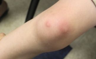
A 5-year old boy presented with these nonpruritic nonsupurrative painful erythematous nodules on his lower extremities. The rash had appeared about 1 week after the onset of a dry hacking cough.

A 5-year old boy presented with these nonpruritic nonsuppurative painful erythematous nodules on his lower extremities. (Figure 1) The rash had appeared about 1 week after the onset of a dry hacking cough. The child had a low-grade fever and joint pain, as well as effusions in his right knee and elbow.
The child had a history of ADHD and seasonal allergies, which were being treated with methylphenidate CR (40 mg/d) and loratadine (5mg/5mL/d). His mother said his daily routine had not changed but due to cooler weather, he had not been playing outdoors. She said he had not been exposed to any new detergents, medications, soaps, or foods.
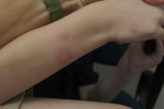
His mother thought the erythema and inflammation might be an allergic reaction to insect bites and had given the child diphenhydramine 5 mL, every 8 hours for the past 48 hours. However, the lesions increased in number and progressed to larger painful, indurated nodules that covered his lower extremities, ankles, and places on his arms and hands. (Figure 2) Lymphadenopathy was noted only in the supraclavicular area.

The child also had coarse basilar breath sounds. A chest x-ray film showed peribronchial changes consistent with bronchitis. A sputum culture was positive for Mycoplasma.
Differentials included erythema multiforme, MRSA, erythema nodosum (EN), and allergic reaction from an unknown irritant or bite.
WHAT’S YOUR DIAGNOSIS?
ANSWER: Erythema Nodosum
Based on the history, morphology, and distribution of the painful nodules, findings of the chest x-ray, and results of sputum culture, a diagnosis of EN, secondary to lower tract respiratory infection, was made. The respiratory infection was treated with azithromycin. Clindamycin provided broad-spectrum coverage until findings were available from joint aspiration and sputum cultures, as well as other laboratory studies. Nonsteroidal anti-inflammatory medications were prescribed until the joint inflammation resolved. Within 24 hours after antibiotic therapy was started, the erythema improved and within 2 weeks, the lesions began to diminish. Joint swelling resolved within a week and aspirates were confirmed aseptic when culture results were returned.
Erythema Nodosum
EN, the most common panninculitis, is thought to be a delayed hypersensitivity response to foreign antigens-often respiratory tract pathogens. It is more common in adults ages 18 to 34 but can occur in children.1 Suspected causes include bacterial infections (streptococcal and mycoplasma), fungal infections, enteropathies, Behcet’s disease, TB, Hodgkin’s disease, and sarcoidosis.2,3 Bacterial infections are the most common cause in children and streptococcal pharyngitis is the most common presentation.3,4
EN typically manifests as painful bilateral pretibial nodules following a febrile illness.1,3 The erythema usually resolves within 1 to 2 days. Nodules most often resolve completely within 6 weeks although they may persist for months, particularly in cases without a clear etiology.3 The lesions are initially firm and tender but can progress to fluctuant abcesses. These rarely suppurate or ulcerate1 which was true in this patient’s case. Nodules most often develop bilaterally on the anterior surfaces of the legs but can occur anywhere and are often associated with joint pain and swelling.1,4 The lesions progress from a red to a purplish to a yellow color resembling the course of a bruise, and then desquamate.1 When synovitis is present it often resolves quickly over weeks but joint pain and stiffness can last months.1 Joint erosion only occurs if there is a secondary infection.
Results of a number of investigations can be useful in making the diagnosis of EN including a complete blood count value, C-reactive protein level, PPD reaction, antistreptolysin titer, erythrocyte sedimentation rate, sputum cultures, skin biopsies, and radiographs of the chest.2 Punch biopsies are not useful. When the diagnosis is unclear, deeper incisional biopsies are required as histopathic changes occur at the subcutaneous level.3,4 Most cases of EN are self limiting and treatment depends on the cause. In this case, results of the sputum culture helped confirm the etiology.
Teaching Points
1. Although Erythema Nodosum is most often a self limiting condition, infectious causes should be ruled out and managed on an individualized basis.
2. A definitive diagnosis can be made with a wide incisional biopsy of presenting nodule(s).
3. The treatment of choice for pain and inflammation is nonsteroidal anti-inflammatory medication along with treatment of any obvious underlying cause.
4. Reassurance and supportive management are useful especially in patients with persistent painful lesions or in disease recurrence.
References:
References:
1. Hebel J, Elston D. Erythema nodosum. WebMD. Accessed March 5, 2011 and available at http://emedicine.medscape.com/article/1081633-overview.
2. GarcÃa-Porrúa C, González-Gay MA, Vázquez-Caruncho M, et al. Erythema nodosum. Etiologic and predictive factors in defined population. Arthritis Rheum. 2000;43:584-92.
3. Requena L, Requena C. Erythema nodosum. Dermatology Online Journal. 2002; 8:4.
4. Schwartz R, Nervi S. Erythema nodosum: a sign of systemic disease. Am Fam Physician. 2007;75:695-700.
