- Clinical Technology
- Adult Immunization
- Hepatology
- Pediatric Immunization
- Screening
- Psychiatry
- Allergy
- Women's Health
- Cardiology
- Pediatrics
- Dermatology
- Endocrinology
- Pain Management
- Gastroenterology
- Infectious Disease
- Obesity Medicine
- Rheumatology
- Nephrology
- Neurology
- Pulmonology
Woman With Transient Year-Round White, Numb, Cold Hands
A woman has white, numb, cold hands after vigorous exercise, emotional stress, and exposure to cold. Is this caused by Buerger disease (thromboangiitis obliterans), acute arterial occlusion, Raynaud disease, or acrocyanosis?
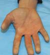
THE CASE: A 24-year-old otherwise healthy woman reports that during the past winter, her hands frequently turned white, numb, and cold. The affected area sometimes included all of the digits on both hands, but more often the reaction was asymmetric and at times involved just half of one digit. The white and numb phase was usually followed by tingling and slight discomfort; then the area turned blue. When the hands were rewarmed, the fingers turned bright red and often throbbed. Episodes occurred after vigorous exercise, severe emotional stress, and during driving, but they typically followed exposure to the cold.

The patient has no history of surgery, medical disorders, or allergies. She does not smoke and her only medication is an oral contraceptive.
Her vital signs are all within normal limits. Pulse oximetry is 100% on room air. The physical examination is unremarkable with the exception of hyperemia of both hands. There is no cyanosis or clubbing. Brachial, radial, and ulnar pulses are strong and symmetric bilaterally. No deformity of the joints or nails, no cutaneous abnormalities, and no sensory deficits are detected. The neurologic examination reveals no proximal (shoulder girdle) weakness. Motor functions of the radial, ulnar, and median nerves are intact.
What is the most likely cause of the patient's symptoms?
- Buerger disease (thromboangiitis obliterans)
- Acute arterial occlusion
- Raynaud disease
- Acrocyanosis
DISCUSSION: This young woman is experiencing transient episodes of digital ischemia associated with Raynaud disease. She exhibits a predictable pattern of digital pallor, cyanosis, and rubor, with eventual return to normal appearance and no signs or symptoms of significant pathology or disease between events.
In patients with Raynaud disease, the digital ischemia is not subtle and may be evoked by cold exposure, emotional stress, and repetitive activity. The initial (ischemic) phase of digital blanching occurs secondary to vasospasm of arteries and arterioles. The cyanotic phase results from engorgement of the venules with deoxygenated blood. Numbness or paresthesia occurs during pallor and cyanosis. After rewarming, vasospasm resolves and blood flows into vessels that are now dilated, which results in a reactive hyperemia. This phase of rubor is often described as painful throbbing.


Raynaud phenomenon is termed Raynaud disease when the cause is idiopathic. About half of patients with Raynaud phenomenon have Raynaud disease. Women are affected 5 times more frequently than men; age at onset is typically between 20 and 40 years. Fingers are affected much more often than toes. When the cause of the vasospasm is identified, or when the vasospasm is associated with another disease, the disorder is referred to as Raynaud phenomenon. Common causes of this condition include scleroderma, systemic lupus erythematosus, dermatomyositis, poliomyelitis, thoracic outlet syndrome, blood dyscrasias, and cryoglobulinemia. Medications that may induce Raynaud phenomenon include ergot preparations, cisplatin, b-agonists, vincristine, and bleomycin.
Patients with Raynaud disease often present with a history of digital ischemia and tricolor changes but no other skin changes (such as ulceration), absent or impaired peripheral pulses, or motor or sensory abnormalities. Some patients exhibit only cool hands and feet. About 10% present with sclerodactyly.
Most cases of Raynaud disease are mild; patients need only to be reassured and advised to dress warmly and avoid cold exposure and cigarette smoking. In patients with severe symptoms, calcium channel blockers (especially diltiazem and nifedipine) reduce the frequency and severity of episodes. Other agents may relieve symptoms slightly in some patients but are not widely used because of adverse effects and limited benefit. These include the a1-adrenergic antagonists prazosin, doxazosin, and terazosin. Reserpine increases blood flow to the digits but is associated with hypotension, nasal congestion, and depression. Surgical sympathectomy is a drastic measure that has only transient effectiveness.
Buerger disease, an inflammatory vascular disorder of the small and medium vessels of the distal upper and lower extremities, is associated with smoking and primarily affects middle-aged men. In the first stage, polymorphonuclear leukocytes (PMNs) invade the vessel walls. Later, mononuclear cells, fibroblasts, and giant cells replace PMNs. Final stages are marked by perivascular fibrosis and recanalization.
Patients with Buerger disease experience claudication of the affected extremity (calf and foot or forearm and hand), Raynaud phenomenon, and migratory superficial venous thrombophlebitis. The physical examination may reveal severe digital ischemia with trophic nail changes, ulceration, and gangrene of the fingertips. Large vessel pulses (brachial and popliteal) are normal; peripheral pulses (radial, ulnar, dorsalis pedis, and posterior tibial) are weak or absent.
The diagnosis may be supported by arteriography but is formally made by biopsy with pathologic evaluation of the vessel walls. There is no effective treatment. Patients must be advised to stop smoking immediately. Surgical bypass of large vessels may be necessary, along with local debridement of ulcers and gangrene or, in extreme cases, amputation of digits and extremities.
Patients with acute arterial occlusion often manifest acute, dramatic occlusion with pallor and ischemia that are not transient or episodic and that progress to a cyanotic and hyperemic phase. Peripheral pulses are often absent. If ischemia is not treated, tissue will become necrotic.
The causes of acute arterial occlusion are thrombus formation and embolism. The most common sources of emboli to extremity arteries are the heart and aorta. Conditions that predispose to thromboembolism are septal defects, atrial fibrillation, myocardial infarction with or without ventricular aneurysm, cardiomyopathy, atrial myxoma, endocarditis, and prosthetic heart valves.
Acute arterial thrombosis occurs most commonly in vessels with atherosclerosis. Trauma to vessels (including arterial punctures and catheter placement) may cause an endothelial disruption with formation of an acute thrombus. Hypercoagulable disorders may also cause thrombosis.
Symptoms of acute occlusion depend on location and duration of the obstruction. Pain, paresthesias, numbness, and coolness develop within 1 hour in the affected extremity. Physical examination reveals reduced or absent pulses, changes in skin color (pallor, cyanosis, or mottling); motor findings (weakness or paralysis); sensory changes (numbness, paresthesias, or pain); and absence of deep tendon reflexes.
Arteriography is useful for diagnosis and for delineating the extent and location of the occlusion. Treatment involves immediate anticoagulation and surgical intervention with thromboembolectomy or arterial bypass grafting. The outcome and prognosis are determined by the length of time elapsed before intervention to return blood flow to ischemic tissue.
Patients with acrocyanosis have arterial vasoconstriction (not vasospasm) of capillaries and venules (not digital arteries), followed by dilation and persistent digital cyanosis. Like Raynaud disease, acrocyanosis more frequently affects women; onset is by age 30 years.
Patients usually present with concern about blue discoloration of their fingers. Physical examination reveals normal peripheral pulses, no motor or sensory abnormalities, and no skin changes, such as ulceration. Arterial oxygenation saturation is normal and there is no evidence of central cyanosis.
Treatment is similar to that for mild Raynaud disease. No medication is recommended for acrocyanosis.
