- Clinical Technology
- Adult Immunization
- Hepatology
- Pediatric Immunization
- Screening
- Psychiatry
- Allergy
- Women's Health
- Cardiology
- Pediatrics
- Dermatology
- Endocrinology
- Pain Management
- Gastroenterology
- Infectious Disease
- Obesity Medicine
- Rheumatology
- Nephrology
- Neurology
- Pulmonology
Unusual Cause of Bilateral Optic Neuritis in a Patient With AIDS
Bilateral retrobulbar optic neuritis developed in a 38-year-old woman with advanced HIV infection. This was secondary to varicella-zoster virus (VZV) infection, confirmed by polymerase chain reaction detection of VZV in the patient's cerebrospinal fluid. There was no evidence of retinitis, and the ocular symptoms preceded the rash. This case illustrates that a new onset of unexplained visual loss resulting from optic neuritis in an HIV-positive patient may be caused by VZV infection. Clinicians should be aware of this unusual manifestation of VZV infection. Prompt recognition and early intervention with antivirals are needed, but it is unclear how much vision can be preserved.
We describe a case of bilateral retrobulbar optic neuritis associated with varicella-zoster virus (VZV) infection in a patient with advanced HIV infection with profound immunosuppression.
CASE REPORT
A 38-year-old woman was transferred to our hospital because of decreased visual acuity, with inability to perceive light in both eyes. She had presented to another hospital complaining of mild to moderate headache for 1 week and of ocular pain and blurred vision in both eyes for 2 days, which rapidly progressed to inability to see light on the day of admission. She had a history of untreated HIV infection for 7 years; her absolute CD4+ cell count was 15/μL and serum HIV RNA level was 169,000 copies/mL.
The physical examination findings were remarkable for dilated pupils with no light perception in either eye. Extraocular movements were intact. Corneas and funduscopic examination findings were normal. Findings on a CT scan of the brain and from cerebrospinal fluid (CSF) analysis also were normal.
After being transferred to our hospital for further evaluation, the patient continued to have moderate to severe headache. One day later, a vesicular rash was noted on the left side of her face (lips, bridge of the nose, and eyelids), and treatment with high-dose acyclovir was started.
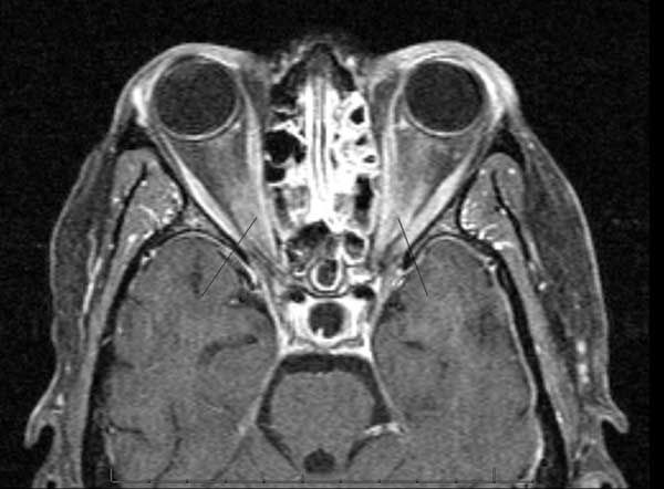
Initial lumbar puncture disclosed a high opening pressure (34 cm H2O) and CSF with 16 white blood cells (91% neutrophils and 9% lymphocytes), normal glucose level, and protein level of 74 mg/dL. Results of a VDRL test, a Cryptococcus antigen test, an acid-fast stain, a Mycobacterium tuberculosis polymerase chain reaction (PCR) assay, and all cultures (viruses, fungi, bacteria, and mycobacteria) were negative. A CT scan of the brain with contrast showed mild atrophy, but an MRI scan showed diffuse enhancement of the bilateral optic nerves (Figure).

Figure. This axial T1-weighted MRI scan after intravenous injection of gadolinium shows marked signal enhancement of both optic nerves, indicating disruption of the blood-nerve barrier and optic neuritis.
The patient’s vesicular rash evolved over the next few days and healed several days later. Her severe headache, which required high doses of narcotics, also improved about 1 week after admission. Eye examination findings were unchanged, and the acyclovir treatment was continued.
A second lumbar puncture, done 10 days after the first, showed normalization of the opening pressure, no cells, normal glucose level, and total protein level of 49 mg/dL. A VDRL test, M tuberculosis PCR assay, Cryptococcus antigen test, and culture results were negative. The results of PCR studies for viruses such as Epstein-Barr virus, herpes simplex virus (HSV) 1 and 2, Cytomegalovirus (CMV), and JC virus (a polyomavirus) were negative, but the test for VZV was positive.
Because of the lack of improvement with specific antiviral therapy, the patient received a short course of high-dose corticosteroids. There were no complications, but her vision did not improve. She was discharged 2 weeks after admission with only modest improvement in vision in her left eye. She did not return to the outpatient clinic for follow-up.
DISCUSSION
We have described a woman with advanced HIV infection with profound immunosuppression in whom bilateral retrobulbar optic neuritis developed as a result of VZV infection. The diagnosis of retrobulbar optic neuritis was based on typical symptoms, such as reduced vision, reduced pupillary contractions, and ocular pain; normal initial anterior segment and funduscopic examination findings; and compatible MRI findings. VZV was implicated as the cause of the optic neuritis on the basis of the posterior development of trigeminal dermatitis; identification of VZV in CSF by qualitative PCR assay; and lack of evidence of alternative causes on the basis of comprehensive serum and CSF test results and radiological findings.
Optic neuritis is defined as the retrobulbar inflammation of the optic nerve.1 This sudden inflammation of the optic nerve leads to edema and destruction of the myelin, although some authors suggest that the inflammation is most frequently caused by demyelination. The inflammation is commonly associated with autoimmune diseases, particularly multiple sclerosis; more than 50% of patients who have optic neuritis will eventually have multiple sclerosis. However, the inflammation may occasionally be caused by infection, such as Lyme disease, tuberculosis, syphilis, hepatitis B virus infection, and infections with the Herpesviridae family (HSV, VZV, and CMV).2
The most common signs and symptoms are acute loss of vision, loss of color vision, pain on movement of the eye, and evidence of optic neuropathy (impaired visual acuity and color vision, visual field loss, and afferent pupillary defects). The pain associated with optic neuritis tends to progress over days. The vision typically improves over 3 to 8 weeks, and most visual recovery occurs within the first 6 months, but it can continue for up to 1 year after the acute event.
Three-fourths of patients have nearly complete return of central vision, as measured by reading the eye chart. However, there is usually some permanent residual haziness, which patients describe as “looking through smoke” even though outlines of objects are sharp. Fifteen percent of patients remain blind in the affected eye. The diagnosis is established on clinical grounds and is complemented by MRI (demonstration of inflammation within the optic nerve).1,2
Varicella-Zoster Virus
VZV, an exclusively human, highly neurotropic alphaherpesvirus, is a member of the Herpesviridae family. It is an enveloped double-stranded DNA virus that establishes lifelong latency in cranial nerve ganglia, dorsal root ganglia, and autonomic ganglia along the entire neuroaxis after the initial infection.3 Primary infection (varicella) is almost always acquired by respiratory droplets and rarely from contact with vesicles/ulcers (zoster).
The initial infection, or varicella, causes an acute febrile illness, usually in childhood, characterized by a widespread vesicular exanthem (chickenpox). CNS complications occur in fewer than 1% of cases and include mild meningitis and acute cerebellitis (the most common neurological abnormality associated with varicella in children and adolescents). Encephalitis and myelitis have been reported as well, but they are quite rare.4
After the acute attack of chickenpox resolves, VZV becomes latent, and the viral DNA may remain extrachromosomal but in a nonin-fectious state. VZV may later reactivate in a ganglion, causing a localized, usually dermatomal, eruption of zoster (shingles). Zoster is known to cause a wide spectrum of neurological conditions, including postherpetic neuralgia, segmental sensory loss or motor paresis from radicular involvement, meningoencephalitis, myelitis, ventriculitis, cerebral vascular occlusion arising from vasculitic involvement, zoster sine herpete (neurological disease without rash), necrotizing retinitis, progressive outer retinal necrosis, and optic neuritis. Almost all cases of optic neuritis described in the literature have been in patients with advanced HIV infection.5-8
Several mechanisms could possibly lead to tissue injury, including cytotoxic or demyelinating effects of the virus itself, vasculitis leading to cord ischemia, or meningitis with release of inflammatory mediators or secondary involvement of penetrating vessels.
Diagnosis
The characteristic rash (either of primary varicella or zoster) usually leads to a clinical diagnosis without the need for additional testing. For definitive diagnosis or for diagnosis of atypical lesions commonly seen in patients with advanced immunosuppression, the following tests are useful: Tzanck test of lesion scrapings, which may demonstrate multinucleated giant cells (60% sensitive for infection with organisms of the Herpesviridae family); direct fluorescent antibody stain of skin biopsy specimen; viral culture (reduced sensitivity if antiviral therapy has been initiated); and VZV PCR assay of lesion exudates or CSF (highly sensitive and specific).9,10
The CSF in VZV myelitis frequently shows an elevated protein level with pleocytosis and normal glucose level, as was seen in this patient. The opening pressure is usually normal or slightly elevated. VZV is rarely isolated on cell culture assays, which have little role in diagnosis.
Rapid diagnosis by PCR amplification of VZV DNA in the CSF is very helpful in diagnosing VZV infection and can provide early support of the diagnosis. There is, however, a diagnostic window for the PCR method in the early phase of the infection, after which viral DNA may disappear in the CSF. During the later stages of CNS infection, testing for VZV IgG antibodies in CSF seems to be the method of choice for diagnosis. Antiviral IgG antibody is not normally found in CSF, so its presence in the absence of red blood cells in the CSF is considered significant.
The diagnosis of a VZV infection in the CNS should not be ruled out in the appropriate clinical setting, even if no VZV DNA or specific IgG antibodies are detected, since it is recognized that none of the available assays have ideal sensitivity.
Treatment
The patient’s age and immune status are the main factors to consider in deciding how to treat zoster, or shingles. In general, the use of oral antivirals (acyclovir, famciclovir, or valacyclovir) is not required in immunocompetent patients younger than 50 years, but there may be some evidence that it will speed healing of the rash. In any patient older than 50 years, antivirals should be used.
In immunocompromised patients, high dosages of intravenous acyclovir (5 to 10 mg/kg 3 times daily for 5 to 7 days) should be used. At any age, trigeminal involvement should also be treated with antiviral drugs. Given evidence of vascular inflammation in many tissue specimens from patients with CNS sequelae of VZV infection, a rationale exists for combining immune suppression with antiviral therapy. Corticosteroids used concomitantly with antivirals are suggested to be of benefit, but rigorous clinical trial evidence is lacking.
References:
References
1.
Balcer LJ. Clinical practice. Optic neuritis.
N Engl J Med
. 2006;354:1273-1280.
2.
Kang PS, Munter FM, Swallow C, et al. Optic neuritis. eMedicine. February 8, 2006.
http://www.emedicine.medscape.com/article/383642-review
.
3.
Nagel MA, Gilden DH. The protean neurologic manifestations of varicella-zoster virus infection.
Cleve Clin J Med
. 2007;74:489-494, 496, 498-499.
4.
De La Blanchardiere A, Rozenberg F, Caumes E, et al. Neurological complications of varicella-zoster virus infection in adults with human immunodeficiency virus infection.
Scand J Infect Dis
. 2000;32:263-269.
5.
Liu JZ, Brown P, Tselis A. Unilateral retrobulbar optic neuritis due to varicella zoster virus in a patient with AIDS: a case report and review of the literature.
J Neurol Sci
. 2005;237:97-101.
6.
Lee MS, Cooney EL, Stoessel KM, Gariano RF. Varicella zoster virus retrobulbar optic neuritis preceding retinitis in patients with acquired immune deficiency syndrome.
Ophthalmology
. 1998;105:467-471.
7.
Friedlander SM, Rahhal FM, Ericson L, et al. Optic neuropathy preceding acute retinal necrosis in acquired immunodeficiency syndrome [published correction appears in
Arch Ophthalmol.
2000;118:543].
Arch Ophthalmol
. 1996;114:1481-1485.
8.
Greven CM, Singh T, Stanton CA, Martin TJ. Optic chiasm, optic nerve, and retinal involvement secondary to varicella-zoster virus.
Arch Ophthalmol
. 2001;119:608-610.
9.
Gilden DH, Mahalingam R, Cohrs RJ, Tyler KL. Herpesvirus infections of the nervous system.
Nat Clin Pract Neurol
. 2007;3:82-94.
10.
Aberle SW, Puchhammer-Stöckl E. Diagnosis of herpesvirus infections of the central nervous system.
J Clin Virol
. 2002;25(suppl 1):S79-S85.
