- Clinical Technology
- Adult Immunization
- Hepatology
- Pediatric Immunization
- Screening
- Psychiatry
- Allergy
- Women's Health
- Cardiology
- Pediatrics
- Dermatology
- Endocrinology
- Pain Management
- Gastroenterology
- Infectious Disease
- Obesity Medicine
- Rheumatology
- Nephrology
- Neurology
- Pulmonology
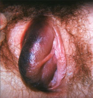
Teenaged Girl With a Unilateral Swollen Purple Labium
A 13-year-old girl is seen because of a genital injury sustained during a fall from her bicycle. Is post-menarchal. Denies any past or present sexual activity, consensual or coercive. Parents report that she has not been ill-adjusted at school and has had no more behavioral issues than her age cohort in recent months.

HISTORY
A 13-year-old girl is seen because of a genital injury sustained during a fall from her bicycle. Is post-menarchal. Denies any past or present sexual activity, consensual or coercive. Parents report that she has not been ill-adjusted at school and has had no more behavioral issues than her age cohort in recent months.
PHYSICAL EXAMINATION
Young adolescent who is appropriately upset due to pain and to fear that her disfigurement will endure. Tachycardia, hypertension but no tachypnea and no fever. No evidence of sexual trauma or of sexually transmitted infection on examination of mouth and anus. Abdominal examination normal.
WHAT'S YOUR DIAGNOSIS?
(Answer on next page.)
ANSWER: VULVAR LABIAL TRAUMA
The striking lesion is a large fresh-appearing hematoma of the right labium minus. One is tempted to say that the labium majus is involved, all the more so because of the unaffected medial diagonal "sash" that connects hematoma to clitoral hood, which one could mistake for the entire ipsilateral labium minus. However, the patient's fingers retract her right labium majus laterally, and the medial delimitation of hair-bearing (labium majus) skin forms the outer margin of a wide normal crescent of skin just lateral to the hematoma. Further complicating anatomical orientation is a dark narrow crescent just medial to the well-lit normal crescent, but this is merely a shadow cast by the hematoma-expanded skin: the lighting falls from the right.
The extent of pubic hair is consistent with developmental age. Skin of the labia majora appears normal. The left labium minus is unremarkable; so is the visualized small portion of the medial surface of right labium majus. We can see the clitoral hood but not the clitoris, nor the urethral meatus. Only the outermost portion of rugate introitus is discernible, abutting a mostly concealed posterior fourchette.
COULD THE STORY BE WRONG?
Every clinician will consider sexual trauma here. Most will note that the lack of lacerations and petechiae does not prove a case for or against rape, nor for or against consensual sexual activity. Detailed discussion of rape examination and collection of evidence exceeds the scope of this column. In one study of prepubescent girls and teenagers, labial hematomas were surprisingly uncommon among victims of sexual assault.1 Consultation with gynecologist as needed, rape counselor, and mental health professional is vital if sexual trauma is considered possible, and often discussion with law enforcement as well. In this instance, the patient, family, and clinician all felt sure that there was no hidden explanation.
NUMEROUS SOURCES, ONE MECHANISM
The notion of a fall from a bicycle with a straddle injury as the excuse for a vulvar hematoma is almost a clich, adduced falsely by patients, parents, husbands, and perpetrators at various times. Yet straddle injuries do frequently cause hematomas of the vulva, and not only from bicycles,2 but also from the boot bindings on snowboards when these are left up with one foot taken out, typically during dismounting from a chairlift.3
The vulva is particularly susceptible to hematoma because of its high vascularity and ready compression against the pubic bone from blunt trauma. Its loose connective tissue offers feeble resistance to spread of extravasated blood. This sequence applies whether the source is intrapartum trauma,4 excessively vigorous consensual intercourse (one recalls injuries sustained in the grip of cocaine), or the appalling damage done by rape, by object rape, and by being kicked in the groin.5,6
PUERPERAL, HEMATOLOGICAL, AND SEXUAL CONNECTIONS
Besides childbirth and rape, what other sources can cause these hematomas? One is abrasion due to an internal sling used for surgical alleviation of urinary incontinence.7 Others have followed hemorrhagic diatheses: leukemia with thrombocytopenia presented with an immense vulvovaginal mass of blood.8 Treatment of essential thrombocythemia was associated with another large vulvar hematoma,9 and in another, the microvasculopathy and coagulopathy of systemic rickettsial infection.10 As to sexual elements, cunnilingus has been reported as a co-cause in the patient with thrombocythemia9 and so has a sexual bite of the vulva compounding acute alcoholic stupor, further complicated by delay in treatment.11 Extravasation from a branch of the internal iliac artery, perhaps due to trauma from chronic urethral self-catheterization in a setting of sensory loss,12 constitutes yet a further rare cause.
NEWER DIAGNOSTICS AND THERAPEUTICS
Many vulvar hematomas are far more disfiguring than the one illustrated. Key features of management include analgesia, rest, icing, and determination that there is not urinary retention from obstruction of the external urethral meatus or distal urethra.12 A subset of cases need surgical interventions, including those hematomas that so enlarge during labor as to block delivery of the fetus!4
Despite the relatively superficial location, there is a role for ultrasound, in particular when continued bleeding occurs at the internal aspect6,8,13 so that there is no dramatic expansion to telegraph brisk, typically arterial, extravasation.2,4
Interventional radiology now plays a role in shutting down such bleeding including via directed microembolization,12,14 without surgery with its attendant risks and complications. Redundancy of arterial supply has prevented skin and mucosal necrosis.14
A short catheter with an inflatable balloon at its proximal end that serves to hold it in place, the Word catheter, allows drainage without sutures in a subset of cases15; this device spares the pain of sutures and their removal in this uncomfortable area, and was also cited in the column on Bartholin abscess management.16
Most women and girls with vulvar hematomas recover completely; persistent dyspareunia or adverse cosmetic effect appears to be extremely rare.
IS THIS TOO MUCH FUSS?
If we recall that underreporting of genital disorders is rampant,3,11 due to embarrassment, to fear of punishment, and to misperceptions that any problem in these parts of the body is likely to be misinterpreted as sexual in origin, we are reminded that expert genital examination is a responsibility for all practitioners.17
Precise localization is often critical to diagnosis. Yet most primary care clinicians can find genital anatomy confusing regardless of patient and personal gender, in part because we all still harbor the wise societal proscription against staring at people's private parts.
Midwives and gynecologists overcome this issue. The rest of us need to work past a subliminal concern that somehow we are violating the patient's privacy. In fact, when we bring the subject into open discussion, it is obvious that we are merely doing our work properly when we scrutinize these parts of the body as we do any others.
But humans are not purely rational. Even knowing that our motives are pure, vestiges of modesty persist. That is good: We deplore callousness about the embarrassment which our routine procedures cause to patients who may never have shown their body to any other person, or only to a parent in childhood and a spouse in adulthood if that. Yet we must override the impulse that would make us a timid examiner. Timidity is an enemy of accurate diagnosis. Nor does it convey respect for patients' personhood: dignified and professional demeanor and choice of words do that job. If anything, the patient who perceives an examiner as hesitant is likely to lose confidence and perhaps therefore to feel more intruded-upon.
So in the expectation that our decency will come across in a powerful and comforting way to the patient, even if nonverbally, we might remind ourselves: "I am looking intently at this person's most private body parts not from an aberrant impulse, nor to violate a social contract or to mortify the patient, but for the most merciful reason, to detect clues to disease so that I can fulfill my fundamental calling to be useful to human beings."
Schneiderman H. Traumatic vulvar hematoma in a teenaged girl, anatomic fine points, and the diversity of causes of vulvar hematomas. CONSULTANT. 2010;50:131-133.
References:
REFERENCES:
1.
McCann J, Miyamoto S, Boyle C, Rogers K. Healing of nonhymenal genital injuries in prepubertal and adolescent girls: a descriptive study.
Pediatrics.
2007;120:1000-1011.
2.
Virgili A, Bianchi A, Mollica G, Corazza M. Serious hematoma of the vulva from a bicycle accident. A case report.
J Reprod Med.
2000;45:662-664.
3.
Kanai M, Osada R, Maruyama K, et al. Warning from Nagano: increase of vulvar hematoma and/or lacerated injury caused by snowboarding.
J Trauma.
2001;50:328-331.
4.
Joy SD, Huddleston JF, McCarthy R. Explosion of a vulvar hematoma during spontaneous vaginal delivery. A case report.
J Reprod Med.
2001;46:856-858.
5.
Shesser R, Schulman D, Smith J. A nonpuerperal traumatic vulvar hematoma.
J Emerg Med.
1986;4:397-399.
6.
Vermesh M, Deppe G, Zbella E. Non-puerperal traumatic vulvar hematoma.
Int J Gynaecol Obstet.
1984;22:217-219.
7.
Richards SR, Balaloski SP. Vulvar hematoma following a transobturator sling (TVT-O).
Int Urogynecol J Pelvic Floor Dysfunct.
2006;17:672-673.
8.
Shivkumar P, Tayade S, Srujana R. Chronic myeloid leukemia presenting as vulvar hematoma.
Int J Gynaecol Obstet.
2008;101:82-83.
9.
Rabinerson D, Fradin Z, Zeidman A, Horowitz E. Vulvar hematoma after cunnilingus in a teenager with essential thrombocythemia: a case report.
J Reprod Med.
2007;52:458-459.
10.
Dietrich JE, Perlman S, Hertweck SP. Post-traumatic vulvar hematoma secondary to coagulopathy caused by rickettsial infection.
J Pediatr Adolesc Gynecol.
2005;18:175-177.
11.
Mathelier AC. Vulvar hematoma secondary to a human bite. A case report.
J Reprod Med.
1987;32:618-619.
12.
Egan E, Dundee P, Lawrentschuk N. Vulvar hematoma secondary to spontaneous rupture of the internal iliac artery: clinical review.
Am J Obstet Gynecol.
2009;200:e17-e18.
13.
Sherer DM, Stimphil R, Hellmann M, et al. Transperineal sonography of a large vulvar hematoma following blunt perineal trauma.
J Clin Ultrasound.
2006;34:309-312.
14.
Kunishima K, Takao H, Kato N, et al. Transarterial embolization of a nonpuerperal traumatic vulvar hematoma.
Radiat Med.
2008;26:168-170.
15.
Mok-Lin EY, Laufer MR. Management of vulvar hematomas: use of a Word catheter.
J Pediatr Adolesc Gynecol.
2009;22:e156-e158.
16.
Schneiderman H. Bartholin gland abscess in the last trimester of pregnancy.
Consultant.
2008;48:543-546.
17.
Schneiderman H. Severe, nonspecific vulvar pruritus with irritation, and other vulvar lesions.
Consultant.
1991;31(1):53-56.
