- Clinical Technology
- Adult Immunization
- Hepatology
- Pediatric Immunization
- Screening
- Psychiatry
- Allergy
- Women's Health
- Cardiology
- Pediatrics
- Dermatology
- Endocrinology
- Pain Management
- Gastroenterology
- Infectious Disease
- Obesity Medicine
- Rheumatology
- Nephrology
- Neurology
- Pulmonology
Takotsubo Syndrome in an African American Woman With Typical Presentation
Also known as “broken-heart syndrome,” Takotsubo syndrome is a stress-induced cardiomyopathy.
A 76-year-old African American woman presented to a local hospital with complaints of left-sided chest heaviness accompanied by shortness of breath and fatigue. Her symptoms, which she said were exacerbated by the hot weather, started the previous afternoon when she encountered a large snake on her front porch. She used a shovel to kill the snake, which required a significant amount of exertion. On a trip to the Laundromat after the incident, the symptoms worsened. The patient denied loss of consciousness but felt physically weak.
The patient was a nonsmoker, and she did not use alcohol. Her past medical history was remarkable for coronary artery disease of the proximal circumflex artery, which was treated successfully with a Taxus drug-eluting stent 5 years previously. She also had hyperlipidemia, type 2 diabetes mellitus, hypertension, and depression. The surgical history was remarkable for a hysterectomy, cholecystectomy, and humeral fracture reduction, from which she had recovered uneventfully.
Memantine and sertraline had been prescribed by her primary care physician. She was also taking clopidogrel, daily aspirin, and antihypertensive and antihyperglycemic medications.
When the patient arrived at the hospital, a troponin T test was done; the second test revealed an elevated troponin T level, at which time a cardiology consultation was requested. The patient’s blood pressure was 131/74 mm Hg; heart rate, 61/min; and respirations, 18/min. She was afebrile. No jugular venous distention was noted, and carotids were 2+ bilaterally without bruits.
The trachea was midline. On auscultation, the heart rate and rhythm were regular, with no clicks, heaves, thrills, or rubs. S1 and S2 were normal. The patient’s lungs were clear, and no masses were detected in her abdomen. There was no clubbing, cyanosis, or edema in the extremities. Findings from the neurologic examination were grossly intact.

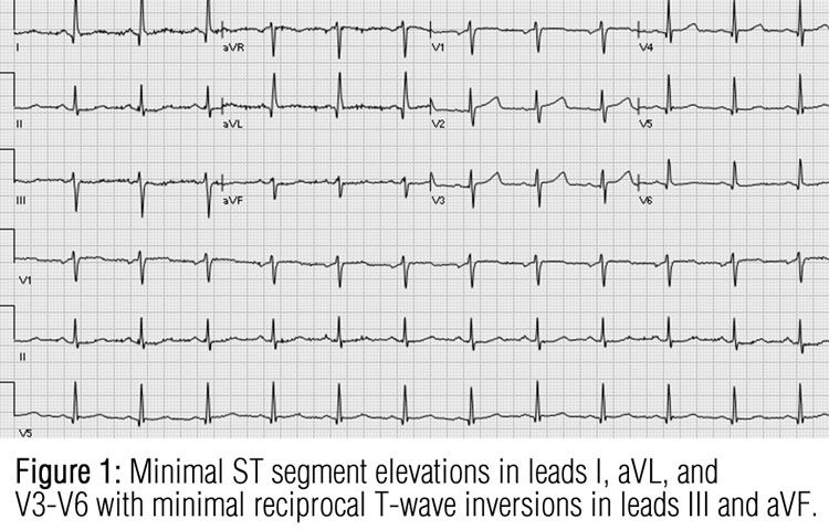
Results of laboratory tests revealed normal levels of blood urea nitrogen, creatinine, low-density lipoprotein cholesterol, magnesium, and phosphorus. However, the patient’s blood glucose level was 196 mg/dL, and her glomerular filtration rate was 48 mL/min. Her troponin T level had been 0.03 ng/mL at presentation; it subsequently rose to 0.21 ng/mL, and finally 0.23 ng/mL. An ECG revealed very minimal ST-segment elevations in leads I, aVL, and V3 through V6 with minimal reciprocal T-wave inversions in leads III and aVF (Figure 1).
On the basis of the history, physical examination findings, laboratory test results, and cardiac studies, a non–ST-segment elevation myocardial infarction caused by a lesion in the left anterior descending or circumflex artery was suspected. The patient was admitted for an echocardiogram and left heart catheterization to fully assess wall motion and coronary anatomy, respectively.

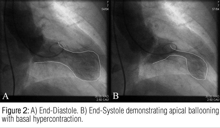
The echocardiogram revealed an akinetic anterior and inferior apex. Mild mitral regurgitation and trace tricuspid regurgitation were also noted; the left ventricular ejection fraction was 45%. Catheterization revealed nonobstructive coronary artery disease, with a 20% tubular stenosis of the mid–right coronary artery and a discrete 30% stenosis of the proximal left anterior descending artery. The circumflex artery and the stent that had been placed previously were patent. Cardiac arteriography with left ventriculogram indicated a large apical aneurysm (ballooning) (Figure 2). These findings led to a diagnosis of stress-induced takotsubo cardiomyopathy. Because the patient had a prolonged QTc, she was monitored for dysrhythmia.
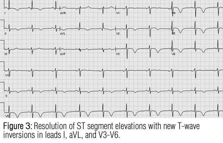
A follow-up ECG demonstrated a resolution of ST-segment elevations, but new T-wave inversions in leads I, aVL, and V3 through V6 were seen (Figure 3). The patient was discharged and directed to continue her usual course of medications. It was recommended that her primary care physician initiate insulin therapy because her current antihyperglycemic regimen no longer appeared to provide adequate control. Her hemoglobin A1C level remained at 9.7% suggesting an average glucose level of 230 mg/dL. The patient was advised to avoid any strenuous activity until her follow-up within 1 to 2 weeks.

Discussion on next page.
Discussion
Takotsubo syndrome, also known as “broken-heart syndrome,” is a stress-induced cardiomyopathy. In Japanese, Tako-tsubo is a word to describe a contraption that is used to catch octopuses.1 This syndrome, which was first described in 1990 by Sato and associates in Japan, usually occurs secondary to emotional stress.2
A 5-patient, single-center study in 2007 by Patel and colleagues3 concluded that African American women may present with atypical symptoms associated with takotsubo syndrome. However, our patient had a typical clinical presentation. In a study by Qaqa and associates,4 African American and non–African American patients were found to have similar presenting symptoms of takotsubo syndrome.
Postmenopausal women are at higher risk for takotsubo syndrome following emotional stress.1,5 This suggests that the disease may be caused by high levels of catecholamines. Patients usually present with symptoms similar to those of coronary artery disease, with ST-segment depression or elevation and T-wave changes.1,6 Most patients will also present with a prolonged QTc and elevation of cardiac enzyme levels.1 Therefore, according to principles of Bayesian analysis, the diagnosis of coronary artery disease and myocardial infarction should be maintained until proved otherwise.2
Apical ballooning on cardiac arteriography with left ventriculogram is typical of takotsubo syndrome and confirms the diagnosis. As seen in Figure 2, apical ballooning and basal hypercontraction can be visualized during systole. Another significant diagnostic finding is apical wall dyskinesia on an echocardiogram.1,2
Although takotsubo syndrome can cause significant morbidity, the treatment is primarily supportive and self-reversal occurs within a few weeks.7 If the patient is hemodynamically stable, β-blockers may be used. In patients with takotsubo syndrome, there is a risk of ventricular thrombus formation as a result of wall akinesia; therefore, anticoagulation may be indicated.8
References:
References
1. Virani SS, Khan AN, Mendoza CE, et al. Takotsubo cardiomyopathy, or broken-heart syndrome. Tex Heart Inst J. 2007;34:76-79.
2. Ibanez B, Benezet-Mazuecos J, Navarro F, Farre J. Takotsubo syndrome: a Bayesian approach to interpreting its pathogenesis. Mayo Clin Proc. 2006;81:732-735.
3. Patel HM, Kantharia BK, Morris DL, Yazdanfar S. Takotsubo syndrome in African-American women with atypical presentations: a single-center experience. Clin Cardiol. 2007;30:14-18.
4. Qaqa A, Daoko J, Jallad N, et al. Takotsubo syndrome in African American vs. non-African American women. West J Emerg Med. 2011;12:218-223.
5. Gianni M, Dentali F, Grandi AM, et al. Apical ballooning syndrome or takotsubo cardiomyopathy: a systematic review. Eur Heart J. 2006;27:1523-1529.
6. Bybee KA, Murphy J, Prasad A, et al. Acute impairment of regional myocardial glucose uptake in the apical ballooning (takotsubo) syndrome. J Nucl Cardiol. 2006;13:244-250.
7. Desmet WJ, Adriaenssens BF, Dens JA. Apical ballooning of the left ventricle: first series in white patients. Heart. 2003;89:1027-1031.
8. Kurisu S, Inoue I, Kawagoe T, Ishihara M, et al. Incidence and treatment of left ventricular apical thrombosis in Tako-tsubo cardiomyopathy. Int J Cardiol. 2011;146:e58-e60.
