- Clinical Technology
- Adult Immunization
- Hepatology
- Pediatric Immunization
- Screening
- Psychiatry
- Allergy
- Women's Health
- Cardiology
- Pediatrics
- Dermatology
- Endocrinology
- Pain Management
- Gastroenterology
- Infectious Disease
- Obesity Medicine
- Rheumatology
- Nephrology
- Neurology
- Pulmonology
Spontaneous Embolization of a Thrombus-in-Transit
Chest pain and dyspnea of acute onset prompted a 49-year-old man to seek urgent medical attention. Two months earlier, he had sustained fractures to the right arm and both ankles after a 25-ft fall. Ten days before presentation, the patient’s rehabilitation physician had discontinued daily enoxaparin because of improved mobility and a presumed decreased risk of thromboembolism.
Chest pain and dyspnea of acute onset prompted a 49-year-old man to seek urgent medical attention. Two months earlier, he had sustained fractures to the right arm and both ankles after a 25-ft fall. Ten days before presentation, the patient’s rehabilitation physician had discontinued daily enoxaparin because of improved mobility and a presumed decreased risk of thromboembolism.

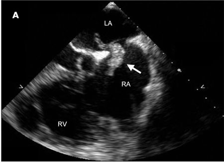
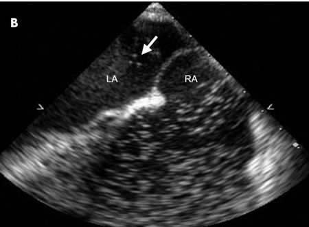
A CT scan of the chest revealed large bilateral pulmonary emboli. A heparin weight-based protocol was started, and transthoracic echocardiography was performed to look for signs of right heart strain. The imaging revealed what appeared to be 2 large echodense structures within the left and right atria. A follow-up transesophageal echocardiography (TEE) revealed a large mobile echogenic structure of 3.6 X 1.5 cm traversing the intra-atrial septum at the level of the foramen ovale, consistent with a thrombus-in-transit (A). During the procedure, the patient began retching and repeated viewing of the intra-atrial septum revealed that the thrombus had completely disappeared. An agitated saline study showed contrast bubbles passing from the right atrium (RA) to the left atrium (LA), confirming the presence of a patent foramen ovale (B).

Although venous thromboemboli are common, paradoxical emboli are relatively rare. The reason is that diagnosis requires an arterial embolus and a venous source in addition to an abnormal communication between the venous and arterial circulation. Even when all criteria are met, almost all cases are diagnosed on a presumptive basis.
Intracardiac thrombi are most often associated with pulmonary emboli and can be visualized on transthoracic or transesophageal echocardiography in as many as 4% to 18% of cases.1 If a patent foramen ovale is present, a thrombus can cross from the right atrial circulation directly into the left atrium. When the thrombus is not attached to any other intracardiac structure, the phenomenon is known as a thrombus-in-transit.2
Following the TEE, the patient regained consciousness and had no shortness of breath, headache, vision changes, or other neurological symptoms. A complete physical examination and close monitoring over the next 24 hours failed to show any new neurological or systemic abnormality. Heparin was continued until an international normalized ratio of 2 to 3 was achieved. Warfarin was then substituted, and the patient was discharged to a rehabilitation facility.
The patient’s lack of systemic symptoms after embolization suggests that the thrombus probably was pushed back into the right atrium and passed into the pulmonary circulation.
References:
REFERENCES:
1.
Goldhaber SZ. Pulmonary embolism.
N Engl J Med.
1998;339:93-104.
2.
Shah DP, Min JK, Raman J, et al. Thrombus-in-transit: two cases and a review of diagnosis and management.
J Am Soc Echocardiogr.
2007;20:1219.e6-1219.e9.
