- Clinical Technology
- Adult Immunization
- Hepatology
- Pediatric Immunization
- Screening
- Psychiatry
- Allergy
- Women's Health
- Cardiology
- Pediatrics
- Dermatology
- Endocrinology
- Pain Management
- Gastroenterology
- Infectious Disease
- Obesity Medicine
- Rheumatology
- Nephrology
- Neurology
- Pulmonology
Snake Bite: A Small Puncture Can Create a Large Problem
An 8-year-old boy from southern Ohio was outside playing when he saw a snake lying in the driveway. The boy picked up the snake to show his father and then dropped it. He picked it up again and was bitten. He sustained a tiny puncture wound to the palmar aspect of his distal left ring finger and a scratch to the distal long finger.
An 8-year-old boy from southern Ohio was outside playing when he saw a snake lying in the driveway. The boy picked up the snake to show his father and then dropped it. He picked it up again and was bitten. He sustained a tiny puncture wound to the palmar aspect of his distal left ring finger and a scratch to the distal long finger.
The boy's father killed the snake and kept it for identification. It was subsequently found to be a copperhead. The family called 911, and the child was taken to the nearest emergency department (ED), which was more than 20 minutes away.
The child had no major medical concerns, no history of surgeries, and no drug allergies. His only daily medication was for attention deficit hyperactivity disorder. His tetanus immunization status was up-to-date (according to the boy's father).
On arrival in the ED, the patient was very tearful and restless and in extreme pain. An intravenous line was placed, and the boy was given normal saline, morphine, cefazolin, and promethazine (Phenergan).
The 2 affected fingers had swelled immediately and moderate edema extended to the palmar crease. The fingers were initially slightly dusky distally, with 3-second capillary refill. The ring finger was the most obviously affected, and had bled immediately after the bite. The medics had applied a sterile gauze dressing with ace wrap and ice pack.
The patient was sedated with midazolam (Versed) and an intradermal test dose of CroFab (antivenin Crotalidae Polyvalent Immune Fab [ovine]) was administered.
The patient showed no initial adverse reaction. An intravenous infusion of CroFab was then started; it consisted of 5 vials of antivenin. The patient's vital signs were normal, and there was no evidence of neurotoxicity. His hand was placed below heart level, and he was secured in a transport helicopter and flown to Columbus Children's Hospital for level I trauma care and a pharmacy-toxicology consult. During the flight, the patient complained of mild itching and dyspnea. He received diphenhydramine (Benadryl) and methylprednisolone. He had no further reaction.

Figure 1

Figure 2
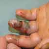
An orthopedic resident, toxicologist, and ED staff examined the patient on his arrival. He complained of severe hand and finger pain along with numbness and tingling. His vital signs were stable. The swelling in his fingers and hand had increased and the dorsal and volar compartments were tense, with clawing of the hand and pain with passive attempt to straighten any of the fingers. Capillary refill exceeded 3 seconds at this point. The ring finger was ecchymotic and dusky (Figure 1). Compartment pressures were measured; thenar compartment pressure was 41 mm Hg; hypothenar, 36 mm Hg; carpal tunnel, 32 mm Hg; and dorsal, 25 and 27 mm Hg. Normal compartment pressures of the hand are 15 mm Hg or lower. If clinical signs and symptoms are present and compartment pressures exceed 30 mm Hg, a compartment syndrome is probably present or imminent.
These measurements and examination results were discussed with the hand surgeon on call and the toxicologist. The plan was to continue close observation while the patient received 5 more vials of CroFab. After 2 hours of observation, repeated examination revealed worsening numbness, swelling, and decreased sensation in all 5 digits. The ring finger was dusky from the proximal interphalangeal joint distally with bullae formation. Repeated measurements of compartment pressures were significantly higher in the thenar (61 mm Hg), hypothenar (45 mm Hg) and carpal tunnel (41 mm Hg) area.
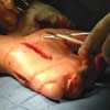
At this point, the hand surgeon decided to operate. Multiple fasciotomies were performed to relieve pressure and prevent necrosis of tissue, muscle, vessels, and nerves (Figure 2).
During the first 3 days of hospitalization, the patient's blood was monitored frequently to watch for alterations in platelet function and red blood cell destruction from the snake toxin.

Figure 3
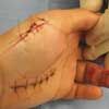
The day after the fasciotomies were performed, the hand surgeon used a vacuum-assisted closure device (VAC; KCI International, San Antonio, Tex) to the open wounds to remove interstitial fluid and debris and to promote wound closure. This negative pressure device was used for 5 days (there were 2 dressing changes during that time). On the sixth postoperative day, the patient returned to the operating room where all of his wounds were closed except for the ring finger fasciotomy site (Figure 3).
The patient received intravenous cefazolin during his 8-day hospitalization. On day 7, he started occupational therapy and range of motion exercises. This continued until 4 weeks after admission. By 6 weeks after the snake bite, the patient had regained normal function of every finger and had no loss of sensation or strength. He was released from care.
SNAKEBITES
There are 8000 venomous bites per year in the United States. There are 12 deaths per year on average, 95% from rattlesnakes.1 Most bites (90%) occur between April and October.1 There are 4 types of venomous snakes in the United States that include the copperhead (pit viper family). Most snake bites are accidental and occur while the victim is doing home improvements, gardening, or recreating. Most bites involve the upper extremity. Fewer than 10% of bites result from curiosity, taunting, or handling.1,2
Pit viper toxin is hemotoxic. It consists of proteins, polypeptides, biogenic amines, and enzymes that cause necrosis and hemolysis. Clinically, reactions can vary from a mild local reaction to life-threatening hemolysis, disseminated intravascular coagulation, or even death (although this is rare). Children suffer more because of their small body size relative to a large venom load. Signs and symptoms of envenomation include local pain within 5 minutes; rapid local swelling, edema, weakness, or numbness/tingling; tachycardia; metallic taste; vomiting; and hypotension and/or shock (which occurs in up to 7% of bites).1-4
Initial treatment includes limiting the patient's extremity activity and using a wide, flat constriction band over the extremity that is not secured tightly (only 20 mm Hg of pressure). The extremity should be immobilized, splinted, and elevated to (but not above) heart level. Suction is rarely used but, if available, it should be applied within 30 minutes of the bite. The device should be an approved model that is correctly applied: otherwise, it will not be effective.1
Stun guns (shock therapy) have been tried in an attempt to neutralize the protein in the venom. However, these have not been found to be effective; in fact, they may exacerbate the clinical situation.5
The patient should be transported to the ED immediately for antivenin therapy, which may be started in the field by medics or during helicopter transportation (if available). Attempts should be made to identify the snake without trying to catch it. Venomous snakes have a distinctly triangular shaped head with elliptical pupils and pits as well as nostrils. They have undivided scales on the underside of their tails, which-except in copperheads-end in a rattle.
Poison control should be contacted as soon as the patient is noted to have a snake bite. All snake bites must be treated as true emergencies, even if the patient appears well at first. The wound(s) should be cleaned, tetanus immunization updated (if appropriate), and intravenous fluids given through 2 large-bore lines. Cardiac, pulse oximetry, and blood pressure monitoring are the norm. Initial laboratory tests include a complete blood cell count with differential (platelets); measurements of electrolytes, glucose, fibrinogen, blood urea nitrogen, creatine, creatinine kinase, and bilirubin levels; coagulation studies; liver function studies; and urinalysis. The patient's blood should be typed and cross-matched.1,3,5
If the clinical examination results are positive, the first step is immediate antivenin therapy.3,4,6,7 There are 2 types of antivenin available in the United States. The antivenin used in this patient's case was a refined and concentrated preparation of serum globulins obtained by fractionating blood from healthy horses immunized with the venom of 4 different species of poisonous snakes. It contains protective substances capable of neutralizing the toxic effects of venoms of multiple snakes. It is only indicated for the treatment of envenomation caused by bites of the crotalids (pit vipers) that are specified (Wyeth Antivenin pharmacy insert). Columbus Children's Hospital stocks CroFab.
Indications for antivenin treatment include:
- Progressive local swelling and ecchymosis.
- Clinically evident coagulation abnormality (low platelet level, increased bleeding time).
- Systemic effects (hypotension, shock, mental status changes).
If none of these is present after a bite, antivenin is not needed. However, the patient should be observed for at least 8 hours.
If antivenin therapy is indicated, the initial dose should be given within the first 4 hours. However, the preparation is effective for up to 24 hours. It can take up to an hour to reconstitute the antivenin from the powder form. The dose is based on the severity of the bite (envenomation grading).1 Children receive the same dose as adults.
After the initial dose is given, the patient is reexamined. The bite is then re-graded. A second or third dose is given as warranted every 2 to 4 hours. Reactions from the antivenin include hypotension, shock, and anaphylaxis. Careful monitoring and extremely close observation are warranted. Our patient was in the pediatric ICU with one-on-one care for over 48 hours.
Surgical treatment for snake bites can include fasciotomies for compartment syndrome. The decision for surgery is based on compartment pressure measurement, not on the clinical examination. In many cases, antivenin therapy alone has been shown to decrease compartment pressures acutely, thereby decreasing the need for surgery.8-11
References:
REFERENCES:
1.
Juckett G, Hancox JG. Venomous snakebites in the United States: management review and update.
Am Fam Physician.
2002;65:1367-1374.
2.
Russell FE, Carlson RW, Wainschel J, Osborne AH. Snake venom poisoning in the United States. Experiences with 550 cases.
JAMA.
1975;233:341-344.
3.
Seger D, Kahn S, Krenzelok EP. Treatment of US crotalidae bites: comparisons of serum and globulin-based polyvalent and antigen-binding fragment anti-venins.
Toxicol Rev.
2005;24:217-227.
4.
Thorson A, Lavonas EJ, Rouse AM, Kerns WP 2nd. Copperhead envenomations in the Carolinas.
J Toxicol Clin Toxicol.
2003;41:29-35.
5.
Dart RC, Gustafson RA. Failure of electric shock treatment for rattlesnake envenomation.
Ann Emerg Med.
1991;20:659-661.
6.
Heard K, O'Malley GF, Dart RC. Antivenin therapy in the Americas.
Drugs.
1999;58:5-15.
7.
Tokish JT, Benjamin J, Walter F. Crotalid envenomation: the southern Arizona experience.
J Orthop Trauma.
2001;15:5-9.
8.
Garfin SR, Castilonia RR, Mubarak SJ, et al. The effect of antivenin on intramuscular pressure elevations induced by rattlesnake venom.
Toxicon.
1985; 23:677-680.
9.
Hall EL. Role of surgical intervention in the management of crotaline snake envenomation.
Ann Emerg Med.
2001;37:175-180.
10.
Shaw BA, Hosalkar HS. Rattlesnake bites in children: antivenin treatment and surgical indications.
J Bone Joint Surg Am.
2002;84:1624-1629.
11.
Whitley RE. Conservative treatment of copperhead snakebites without antivenin.
J Trauma.
1996; 41:219-221.
