

- Clinical Technology
- Adult Immunization
- Hepatology
- Pediatric Immunization
- Screening
- Psychiatry
- Allergy
- Women's Health
- Cardiology
- Pediatrics
- Dermatology
- Endocrinology
- Pain Management
- Gastroenterology
- Infectious Disease
- Obesity Medicine
- Rheumatology
- Nephrology
- Neurology
- Pulmonology
Skin Carcinomas: The Isthmus Excision Technique
Two skin carcinomas that are in close proximity can be removed in a single excision.
Two skin carcinomas that are in close proximity can be removed in a single excision.
I call this technique the “isthmus excision” (Figures 1, 2, 3, 4, and 5).


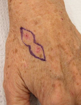
Figure 1 – The excisional isthmus is drawn out.


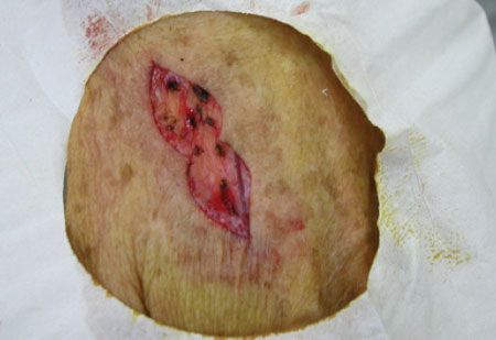
Figure 2 – Both skin carcinomas are excised.


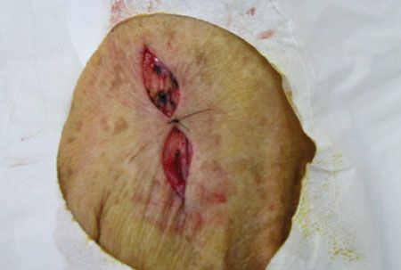
Figure 3 – The isthmus is sutured together. Note the creation of 2 ellipses.
In this instance, the isthmus is a narrow anatomical part connecting 2 larger structures. Instead of 1 large excision, in which the widest part of the margin would be under stress, this technique joins 2 elliptical structures with a narrow isthmus of skin. The isthmus is sutured together, creating 2 separate ellipses that now can be closed into 1 linear incision. The result lessens the possibility of incisional dehiscence.


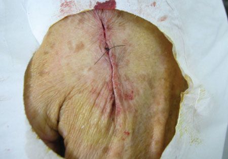
Figure 4 – Subcutaneous closure of the ellipses is shown.


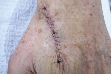
Figure 5 – Final nylon closure is achieved. An interlocking suture technique is shown here, but simple interrupted sutures may be used.
Finally, it is important to alert the pathologist that 2 lesions have been incorporated into 1 excision.
The specimen should be tagged to facilitate anatomical orientation.
-Terrell R. Wallace, Jr, PA-C
Warner Robins, Ga


