- Clinical Technology
- Adult Immunization
- Hepatology
- Pediatric Immunization
- Screening
- Psychiatry
- Allergy
- Women's Health
- Cardiology
- Pediatrics
- Dermatology
- Endocrinology
- Pain Management
- Gastroenterology
- Infectious Disease
- Obesity Medicine
- Rheumatology
- Nephrology
- Neurology
- Pulmonology
Shoulder Fracture-Dislocation in an Elderly Woman: Would You Operate?
An 89-year-old woman, who has long lived on the special care (dementia) unit of a nursing home because of advanced Alzheimer disease, is seen to assess possible injuries after a fall. Many prior falls have been ascribed to her lack of safety awareness in negotiating the environment, rather than to neuromuscular, sensory, or cerebellar deficits.

HISTORY
An 89-year-old woman, who has long lived on the special care (dementia) unit of a nursing home because of advanced Alzheimer disease, is seen to assess possible injuries after a fall. Many prior falls have been ascribed to her lack of safety awareness in negotiating the environment, rather than to neuromuscular, sensory, or cerebellar deficits. None of the falls were syncopal, so arrhythmia is not a consideration. She does not have orthostatic hypotension.
The fall was not observed. Staff quickly responded to the patient's cries and found her lying on the floor. Her right arm was pinned beneath her trunk but was not bent backward or distorted by the weight of her body. No injuries elsewhere. Her mental status and behavior are unchanged from her very impaired baseline.
PHYSICAL EXAMINATION
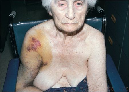
Aged woman who is totally uncooperative with examination, as she has been during each scheduled medical visit for the past several years. Vital signs are normal. She shouts, "Get away from me!" when one touches any part of her body, not just the freshly bruised area shown. She does not appear agitated when observed from afar, although quickly becomes distressed when one asks to examine her or tries, however gently and non-threateningly, to do so.
"WHAT'S YOUR DIAGNOSIS?"
ANSWER: FRACTURE-DISLOCATION OF RIGHT SHOULDER

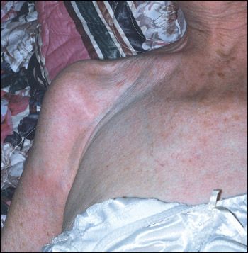
Figure – Three years after the fracture, the axis of the arm shows only trace angulation marking the old fracture site. Extensive hematoma has long since resolved. Linear white streaks at the shoulder suggest surgical scars, but the joint and the humerus were never operated on. The breast and axilla are also now ecchymosis-free.
The image is strikingly upsetting, both because of the deformity of the upper humerus and because the patient's expression-mouth and eyes both-appears so very grim and is easy to overinterpret as indicating extreme pain. In fact, she was not disconsolate, but the camera caught her worst moment. Curiously, the severe bruising appears very deep distally, so that there is an indigo-to-green discoloration "from within," whereas red cells are near the surface in the most proximal portion, despite a paucity of surface abrasion, and also on the medial portion of the anterior axilla and onto the upper outermost part of the breast. Marked yellow-brown discoloration surrounding the purple areas suggests either breakdown of red cells or staining, principally by serum with lesser numbers of erythrocytes.
Based on the extent of the ecchymosis and the deformity that displaces the upper part of the humerus laterally from the main part of the upper arm, a fracture-dislocation was strongly suspected. Radiographs confirmed an acute fracture of the proximal humerus, also known as a surgical neck fracture or fracture of the greater tuberosity (of the humerus).
Orthopedic consultation was undertaken, but the joint decision by geriatrician, family, and surgeon favored conservative management as opposed to surgical procedure. The measures employed yielded a functionally satisfactory result (Figure).
WHY NOT OPERATE?
The idea of forgoing surgery except in extremis or in the setting of prohibitive operative risk will trouble some clinicians. Others may believe that the axis of the arm ought to be set so as to knit in perfect alignment. However, the decision against operating was reached with careful consideration of individual wishes and outcomes: the patient's deeply loving and involved daughters had reminded staff, electively, long before this crisis, that her advance directives stipulated no surgery under any circumstances.
That decision actually made very good sense medically: operation would require time in a hospital, where demented elderly patients experience disorientation, great anxiety, anguish, misery, and delirium, as well as the more conventionally physical problems of a heightened incidence of pressure sores (decubitus ulcers), nosocomial infection, fecal impaction, pulmonary emboli, and death.1 This does not impugn acute hospital staff, who are caring and hardworking. Rather, it reflects empiric observation and the knowledge that the hospital is intrinsically a poor match with the needs of the elderly person, especially the frail aged person and, most of all, the demented, frail older person.
Why do so many untoward complications occur in most acute hospitals? Patients like this one are frail.2 They thrive on utter familiarity of environment and routine, something that cannot be replicated even by a week in one place-which is a long stay in an acute hospital. Nurses, certified nursing assistants, and physicians have to know a demented person for a long time to recognize when something is amiss against a profoundly confusing background of multiple functional impairments and communication deficits. Spontaneous symptom reporting among demented persons is poor to nil. Furthermore, the symptoms and presentation of disease in this group often differ from the expected patterns.3-9 To respond in a timely fashion to a new problem, one has to know the baseline degree of dementia and behavioral aberration, so that one neither overreacts to behavior that is usual for the patient nor fails to spot indicators of new trouble.
WHAT HAPPENED NEXT
All were advised that a satisfactory outcome was obtainable with conservative measures. Thus the patient was spared the expected adverse effects of being uprooted-and all who knew her concurred with the daughters' decision on her behalf. Opiates were used as needed for effective and nontoxic pain control acutely, and were soon tapered to zero; the patient experienced no lasting discomfort. The bone knit, as bones usually will, even without surgery. The patient regained the same limited use of the limb for eating, dressing with assistance, and recreation that she had had before the fracture; the limiting factor was her cognitive status, for she was markedly apraxic, and the fracture contribution was actually very slight.
Although one could be concerned that laziness or conflicting motives might underlie the choice against operation, such a decision actually entails more and harder work for the primary care clinician: time is spent, appropriately, speaking at some length with the parties, thinking about the choice, and revisiting it (perhaps with a sleepless night of self-doubt thrown in). If the medical reasoning and the emotional tone of both family and health care team have been sound, the decision should stand up to any critique, including the harshest of all-self-critique. There are startlingly few reports in the literature on what to avoid in the care of the most demented persons.10 When one helps a family to reach a good decision in an emotionally difficult situation such as this one, the tenor of the relationship with the patient and the family is much more often enhanced, not diminished, by the process and the choice. Such was the case with this patient, who lived several years longer and died of an unrelated problem that posed its own hard choices; these will be the subject of a future "What's Your Diagnosis?" column.
BLEEDING
There is almost no such thing as a medical condition that clears up uneventfully in demented elderly patients. The immense size of our patient's hematoma made us wonder if a large vessel had been avulsed in the fall, but there was no feature of arterial or venous compromise. The patient had no coagulopathy, antithrombotic medication, or thrombocytopenia that would predispose her to excessive bleeding. Yet the hematoma kept enlarging over the first several days; concern about potential compartment syndrome raised the issue of fasciotomy. Mercifully, the hematoma peaked and began to recede just as these discussions were in progress.
The source of bleeding was capillaries or venules. Bleeding was aggravated by continued movement. This woman moved constantly when not confined by an immobilizing device. She was unable to learn, even after a hundred reminders in both words and action, that each movement would produce pain. The fracture, on radiograph, was not jagged. The broken ends, although displaced, did not seem to rake the adjacent soft tissues. In a different patient, one might have undertaken phlebography, arteriography, or magnetic resonance angiography to look for a rent vessel. We were unwilling to subject this patient to a purely diagnostic procedure that could not enhance management, since the daughters remained consistent: they said they would not authorize operation even if a major vessel were damaged.
FRACTURE-DISLOCATION AND INSUFFICIENCY FRACTURE
Many readers will make the diagnosis on sight. Others may raise the excellent differential diagnosis of soft tissue bleeding, which produces a purely superficial deformity that mimics the effect of fracture. If that occurs, however, it surely cannot be diagnosed until one has a radiograph in hand that shows no fracture.
Neoplasm with traumatic bleeding is a remote prospect, although trauma can unmask bone cancer- a presentation sometimes noted in osteosarcoma and Ewing sarcoma in children. A primary bone tumor would be statistically unlikely here (but see below). The substrate of this fracture is clearly minor trauma in the setting of osteoporosis, although osteoporotic breaks are far more common in weight-bearing bones, namely, the femur and the spine, where they yield disastrous hip fracture and painful compression fractures, respectively. However, the concept of insufficiency fracture11-16 has recently gained more recognition in the literature: these are akin to the old pathologic fracture in that they cannot be explained by the physical forces brought to bear. In an insufficiency fracture, however, the underlying abnormality of the bone is not malignant neoplasia or granulomatous disease, but simple osteoporosis (or osteomalacia, or Paget disease of bone) with attenuation of weight-bearing and shear-resistant biophysical properties. Insufficiency fractures have been reported not only at the expected sites of greatest stress but also in the sternum and almost everywhere in the body.
PHYSICAL FINDINGS AND DIFFERENTIAL DIAGNOSIS
The physical signs in fracture-dislocation of the shoulder are sudden pain, edema, and deformity, often with substantial hematoma, almost always after trauma. A fracture-dislocation after only mild trauma would make one think, concerning this patient, of any or all of these:
Two standard textbooks of orthopedic examination describe no further specific signs to seek in diagnosing shoulder fracture, humeral fracture, or fracture-dislocation.17,18 Numerous signs for instability of the shoulder joint exist,17 but they would be impossible to elicit in this uncooperative patient, and the diagnosis can be made without them. The usual setting for fracture of the proximal humerus is osteoporosis. Only some 20% of such fractures show significant displacement. Pain is expected with even minor movement.
A report of loss of feeling in the arm would raise the question of concomitant injury to the brachial plexus, and had our patient exhibited features of such injury, it would have been that much more distressing to stick with a conservative plan of action; likewise if pallor of the forearm and hand led to discovery of a lost radial pulse, implying injury of the axillary artery. Her right radial pulse was one of the few things that she did permit us to check, and it was normal; rapid neurologic examination, largely done surreptitiously, showed no deficit apart from refusal to move the right arm, which could be attributed to pain. Sensory testing was inconclusive: when the patient saw an examiner approaching to touch the limb, screams followed long before contact could be made.
COMORBIDITIES: FURTHER OBSERVATIONS
This patient's photograph illustrates how the camera can mislead: the aperture of her left eye looks wider than that of the right. Is the right side of her face less mobile than the left? In person, there was no such asymmetry. Observation over time also revealed no such abnormalities. Solar damage, heavy wrinkling, and minor procedures to remove early skin cancers are the only history discernible from this patient's face.
The right breast looked more out-deviated than the left. Here the camera spoke true, but the cause was not primary breast disease or asymmetry. Rather, this apparent asymmetry arose from the breast's having been pressed back for a moment by the edematous, blood-filled right arm.
As to the arm, the enlargement extended to the elbow and below, such that in the Index photograph, one notes discoloration from the bleeding even to the distalmost area within the image. The superior and proximal extent of the initial right arm deformity was not evident until I looked at an "after" photograph; then I could observe more clearly the hollow of the shoulder and the acromion. The spread of the blood, and its serum, went downward gravitationally, since the hand is usually below shoulder level regardless of body position; and the patient's sarcopenic tissue provided less fascial resistance to spread than one would expect in a younger and more vigorous host. Doubtless the quality of the tissue also contributed to poor local tamponade of any ruptured capillaries or venules, hence much more bleeding than expected.
Finally, a newer insight: it had been thought for a long time that brown-yellow color in a bruise meant it must be more than a day old, but a study with superior methodology19 showed that this could occur acutely, presumably from layering of the blood content, such that the browner and yellower parts held fewer red cells and more plasma.
Schneiderman H. Fracture-dislocation of right shoulder in a woman with severe dementia, and the new concept of insufficiency fracture. CONSULTANT. 2006;46:1373-1381.
References:
REFERENCES:
1.
Creditor MC. Hazards of hospitalization of the elderly.
Ann Intern Med.
1993; 118:219-223.
2.
Wilson JF. Frailty--and its dangerous effects--might be preventable.
Ann Intern Med.
2004;141:489-492.
3.
Bayer AJ, Chadha JS, Farag RR, et al. Changing presentation of myocardial infarction with increasing old age.
J Am Geriatr Soc.
1986;34:263-266.
4.
Gambert SR. Atypical presentation of diabetes mellitus in the elderly.
Clin Geriatr Med.
1990;6:721-729.
5.
Hodkinson HM. Non-specific presentation of illness.
Br Med J.
1973;4:94-96.
6.
Berger RG, Levitin PM. Febrile presentation of calcium pyrophosphate dihydrate deposition disease.
J Rheumatol.
1988;15:642-643.
7.
Perry A, Linzer M. Neurologic presentation of a non-neurologic disorder.
Geriatrics.
1989;44:113-154.
8.
Schneiderman H, Dana MF. A serpiginous blood blister and "scabies bites": a confusing presentation of bullous pemphigoid.
Consultant.
2004;44:449-457.
9.
Bhasin N, Berridge DC, Scott DJ, et al. Penile ulcer: an unusual presentation of cholesterol emboli.
Eur J Vasc Endovasc Surg.
2004;27:447-448.
10.
Schneiderman H. Flexion contractures and the physical examination of patients with advanced dementia.
Consultant.
2002;42:1382-1387.
11.
Samdani S. Pelvic insufficiency fractures.
J Am Geriatr Soc.
2004;52:854-855.
12.
Tokuya S, Kusumi T, Yamamoto T, et al. Subchondral insufficiency fracture of the humeral head and glenoid resulting in rapidly destructive arthrosis: a case report.
J Shoulder Elbow Surg.
2004;13:86-89.
13.
Min JK, Sung MS. Insufficiency fractures of the sternum.
Scand J Rheumatol.
2003;32:179-180.
14.
Soubrier M, Dubost JJ, Boisgard S, et al. Insufficiency fracture. A survey of 60 cases and review of the literature.
Joint Bone Spine.
2003;70:209-218.
15.
Iwamoto J, Takeda T. Insufficiency fracture of the femoral neck during osteoporosis treatment: a case report.
J Orthop Sci.
2002;7:707-712.
16.
Wild A, Jaeger M, Haak H, Mehdian SH. Sacral insufficiency fracture, an unsuspected cause of low-back pain in elderly women.
Arch Orthop Trauma Surg.
2002;122:58-60.
17.
Snider RK, ed.
Essentials of Musculoskeletal Care.
Rosemont, Ill: American Academy of Orthopaedic Surgeons; 1997:30-31, 70-80, and especially 77, 91-93, 101-103, 114.
18.
McRae R.
Clinical Orthopaedic Examination.
4th ed. New York: Churchill Livingstone; 1997:41-60.
19.
Mosqueda L, Burnight K, Liao S. The life cycle of bruises in older adults.
J Am Geriatr Soc.
2005;53:1339-1343.
