- Clinical Technology
- Adult Immunization
- Hepatology
- Pediatric Immunization
- Screening
- Psychiatry
- Allergy
- Women's Health
- Cardiology
- Pediatrics
- Dermatology
- Endocrinology
- Pain Management
- Gastroenterology
- Infectious Disease
- Obesity Medicine
- Rheumatology
- Nephrology
- Neurology
- Pulmonology
Severe Psoriasis in Advanced HIV Infection
A 50-year-old African American man with HIV infection had a CD4+ T-cell count of 18/μL (1%), CD8+ cell count of 1035/μL (69%), and CD4:CD8 ratio of 0.01 at the time of diagnosis. He had multiple erythematosquamous skin lesions over his forehead, face, chest, back, and extremities
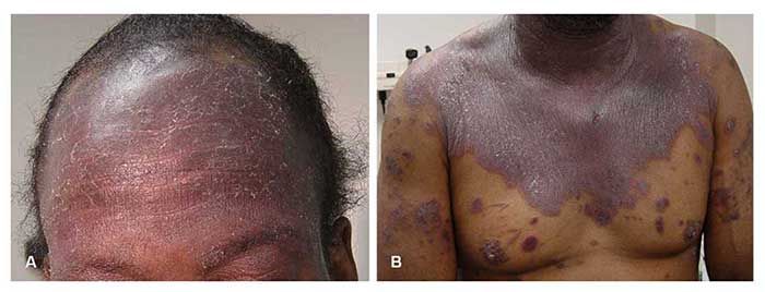
A 50-year-old African American man with HIV infection had a CD4+ T-cell count of 18/μL (1%), CD8+ cell count of 1035/μL (69%), and CD4:CD8 ratio of 0.01 at the time of diagnosis. He had multiple erythematosquamous skin lesions over his forehead, face, chest, back, and extremities (Figure 1). The lesions were characteristic of psoriasis, which was later confirmed with a skin biopsy. Repeated laboratory tests during the initial visit to the HIV/AIDS clinic showed a CD4+ T-cell count of 15/μL (1%); CD8+ cells, 83%; CD4:CD8 ratio, 0.01; and HIV-1 RNA level, greater than 750,000 copies/mL.

Figure 1. The patient had hyperkeratotic plaque and superficial scaling with reddish-based lesions on his forehead (A) and multiple plaques on his chest (B).
The patient began therapy for both psoriasis and HIV infection. His regimen for psoriasis included hydroxyzine 25 mg orally 3 times a day, triamcinolone ointment 0.1% twice daily, and hydrocortisone lotion 1% 3 times daily. His antiretroviral regimen consisted of coformulated lopinavir/ritonavir and zidovudine/lamivudine. Additional medications included azithromycin as prophylaxis against Mycobacterium avium-intracellulare infection and sulfamethoxazole/trimethoprim double-strength as prophylaxis against Pneumocystis jiroveci pneumonia and toxoplasmosis.
Eight weeks after initiation of antiretroviral therapy, the CD4+ T-cell count was 76/μL (5%); CD8+ cells, 75%; CD4:CD8 ratio, 0.07; and HIV RNA level, less than 200 copies/mL. The patient reported good adherence to the prescribed regimen and no adverse effects. There was a marked improvement of the psoriatic lesions.

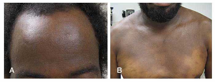
Figure 2. Resolution of the lesions can be seen on the patient's forehead (A) and chest (B).
Three months later, the CD4+ T-cell count had increased to 158/μL (6%); CD8+ cells, 67%; CD4:CD8 ratio, 0.09; and HIV RNA level, less than 50 copies/mL. There was further improvement of the skin lesions, with no visible raised lesions. Hyperpigmentation replaced the old lesions (Figure 2). In his last visit to our clinic, 4 years after initiation of antiretroviral therapy, the CD4+ T-cell count was 305/μL (12%); CD8+ cells, 55%; CD4:CD8 ratio, 0.22; and HIV RNA level, less than 48 copies/mL. There was no clinical evidence of the psoriatic lesions.
Psoriasis is a common chronic inflammatory skin disease with a worldwide prevalence rate of 0.6% to 4.8%. A population-based study in the United States estimated that the prevalence of psoriasis was 1.4% to 4.6% in Caucasians and 0.45% to 0.7% in African Americans.1 The prevalence of psoriasis in HIV-infected persons is not higher than in the general population.2-5 Nevertheless, psoriasis may appear for the first time or preexisting psoriasis may be exacerbated during HIV infection. Psoriasis reportedly becomes more severe with progression of HIV disease6 but may remit in the terminal phase.
The pathogenesis of HIV-associated psoriasis is poorly understood.7 There are several mechanisms proposed to explain the paradoxical exacerbation of psoriasis in advanced HIV infection, including disruption of the skin immune system,3,4 immunodysregulation,5,8,9 superantigens resulting from bacterial or viral infections,1,4,10 genetic factors,10,11 autoimmunity caused by retroviruses,10,11 and a direct role of HIV.2,8,12
As in non–HIV-infected persons, there are several treatment options for psoriasis in HIV-infected persons, including topical therapy, phototherapy, systemic therapy, and tumor necrosis factor (TNF) blockers.7 The clinical improvement seen in patients with severe psoriasis after the institution of antiretroviral therapy suggests benefit from immune reconstitution. This supports the role of immunodysregulation in the pathogenesis of HIV-associated psoriasis. Advanced HIV infection represents a unique opportunity for studying the role of CD8+ memory T cells, TNF-α, interferon, and other immune-mediated mechanisms in the development of psoriasis in non–HIV-infected persons.
References:
References1. Schön MP, Boehncke WH. Psoriasis. N Engl J Med. 2005;352:1899-1912.
2. Fife DJ, Waller JM, Jeffes EW, Koo JY. Unraveling the paradoxes of HIV-associated psoriasis: a review of T-cell subsets and cytokine profiles. Dermatol Online J. 2007;13:4.
3. Maurer TA. Dermatologic manifestations of HIV infection. Top HIV Med. 2005;13:149-154.
4. Namazi MR. Paradoxical exacerbation of psoriasis in AIDS: proposed explanations including the potential roles of substance P and gram-negative bacteria. Autoimmunity. 2004;37:67-71.
5. Mallon E, Bunker CB. HIV-associated psoriasis. AIDS Patient Care STDS. 2000;14:239-246.
6. Mamkin I, Mamkin A, Ramanan SV. HIV-associated psoriasis. Lancet Infect Dis. 2007;7:496.
7. Patel RV, Weinberg JM. Psoriasis in the patient with human immunodeficiency virus, Part 2: review of treatment. Cutis. 2008;82:202-210
8. Vittorio Luigi De Socio G, Simonetti S, Stagni G. Clinical improvement of psoriasis in an AIDS patient effectively treated with combination antiretroviral therapy. Scand J Infect Dis. 2006;38:74-75.
9. Anderson RB, Holman JR. Psoriasis: update on therapeutic choices. Consultant. 2007;47:461-468
10. Mallon E, Young D, Bunce M, et al. HLA-Cw*0602 and HIV-associated psoriasis. Br J Dermatol. 1998;139:527-533.
11. Mallon E. Retroviruses and psoriasis. Curr Opin Infect Dis. 2000;13:103-107
12. Fischer T, Schwörer H, Vente C, et al. Clinical improvement of HIV-associated psoriasis parallels a reduction of HIV viral load induced by effective antiretroviral therapy. AIDS. 1999;13:628-629.
