- Clinical Technology
- Adult Immunization
- Hepatology
- Pediatric Immunization
- Screening
- Psychiatry
- Allergy
- Women's Health
- Cardiology
- Pediatrics
- Dermatology
- Endocrinology
- Pain Management
- Gastroenterology
- Infectious Disease
- Obesity Medicine
- Rheumatology
- Nephrology
- Neurology
- Pulmonology
Seeing HIV/AIDS in Primary Care: A Photo Quiz
Better understanding of this complex condition can lead to better patient care and prevention. This week’s photo quiz offers several presentations to test your knowledge.
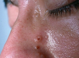
Question 1:
A 38-year-old openly homosexual man presented with cough and multiple asymptomatic facial lesions. Physical examination showed many small, flesh-colored, umbilicated papules. Hospital screening labs revealed a positive HIV serology and gross lymphopenia. A skin biopsy of a facial papule confirmed the diagnosis, a defining opportunistic infection for AIDS.
NEXT QUESTION »
For the discussion, click here.
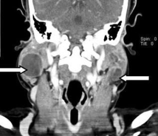
Question 2:
A 36-year-old woman with HIV infection had acute right-sided neck pain that progressed to swelling above the mandible. Ultrasound showed bilateral, anechoic, and primarily cystic lesions in the parotid glands. A CT scan detected a 3-cm, well-circumscribed, and homogeneously hypodense cystic lesion in the right parotid gland and a smaller, 1.2-cm multicystic lesion in the left (arrows). There were subcentimeter lymph nodes in the cervical and supraclavicular chains bilaterally. The condition can be a diagnostic indicator of underlying HIV infection.
NEXT QUESTION »
For the discussion, click here.
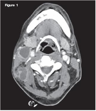
Question 3:
A 38-year-old HIV-infected man was admitted to the hospital after 1 month of painful right neck swelling and 1 week of dysphagia. A CT scan of the neck showed multiple low-density masses with rim enhancement in the right side. A CT scan of the orbits demonstrated abnormal soft tissue involving the right pterygopalatine fossa. Culture confirmed the presence of Aspergillus fumigatus. The diagnosis was invasive aspergillosis.
NEXT QUESTION »
For the discussion, click here.
For the answer, click here.
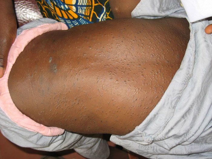
Question 4:
A recently immigrated 18-month-old boy from Nigeria, whose parents remained there ill with AIDS, presented with a fever and a widespread presumably itchy eruption. Considering the family history, tests for HIV infection were obtained; the results were positive. The boy was observed vigorously scratching at rash-filled areas, establishing that the eruption was pruritic.
NEXT QUESTION »
For the discussion, click here.
Question 5:
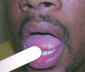
A 30-year-old man with a 15 pack-year smoking history presented for a follow-up evaluation of an asymptomatic whitish lesion on the tongue that had not responded to oral therapy. Multiple, nontender, whitish, vertical striations were noted along the right lateral edge of the lesion. A presumptive clinical diagnosis was made of the classic intraoral lesion of HIV/AIDS.
NEXT QUESTION »
For the discussion, click here.
Question 6:
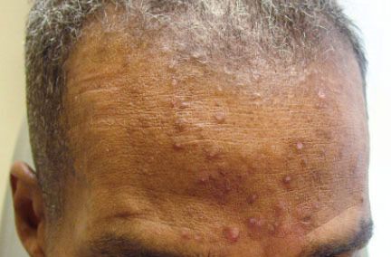
An HIV-positive, 48-year-old man presented with new-onset acne-like, pruritic lesions on his face. A biopsy specimen was obtained from his forehead. He had eosinophilic pustular folliculitis (EPF).
NEXT QUESTION »
For the discussion, click here.
Question 7:
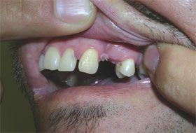
HIV-positive persons typically have a higher rate of human papillomavirus (HPV) than do HIV-negative persons. A 34-year-old man appeared healthy, but examination of the oral mucosa revealed a friable, filiform, digitate lesion on the upper gum. Present for several months, the lesion bled occasionally when he brushed his teeth. Results of HIV testing were positive. A clinical diagnosis of HPV infection was made.
NEXT QUESTION »
For the discussion, click here.
Question 8:
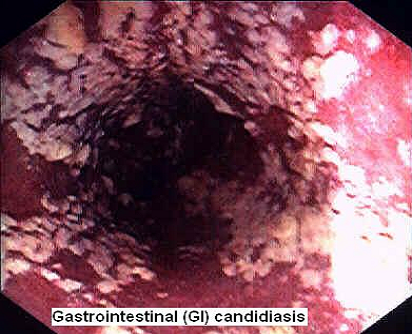
A 47-year-old HIV-positive man presented with severe thrush, profound dysphagia, and a 30-lb weight loss. He underwent an upper endoscopy with biopsies. Severe esophagitis was seen; biopsy specimens revealed unspeciated Candida. Advanced HIV disease made him vulnerable to the dramatic growth of Candida in his esophagus. He had oroesophageal candidiasis.
ANSWER KEY »
For the discussion, click here.
ANSWER KEY:
Question 1. B
Question 2. C
Question 3. A
Question 4. A
Question 5. B
Question 6. E
Question 7. D
Question 8. E
Common Side Effects of Antiretroviral Therapy in HIV Infection
February 7th 2013What are some of the more common side effects of antiretroviral therapy, and what can the primary care physician do to help manage these effects? In this podcast, infectious disease expert Rodger MacArthur, MD, offers insights and points readers to updated comprehensive guidelines.
