- Clinical Technology
- Adult Immunization
- Hepatology
- Pediatric Immunization
- Screening
- Psychiatry
- Allergy
- Women's Health
- Cardiology
- Pediatrics
- Dermatology
- Endocrinology
- Pain Management
- Gastroenterology
- Infectious Disease
- Obesity Medicine
- Rheumatology
- Nephrology
- Neurology
- Pulmonology
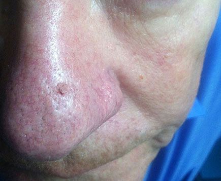
Seborrheic Keratosis Removed Using Shave Biopsy
Histopathology showed this neoplasm to be a seborrheic keratosis: it might well have been a hypertrophic actinic keratosis, melanoma, or superficial basal or squamous cell carcinoma.

A 69-year-old man complained about an asymptomatic “spot” on his nose that had been present for at least 3 months. His past medical history included innumerable facial actinic keratoses.
Key points: Physical examination disclosed a solitary, minimally elevated, 3.7-mm patch on the anterior nose. The area demonstrated a different texture than the rest of the nose and was also characterized by a small eccentric focus of crescent-shaped pigmentation.
Treatment: Because of the small lesion size, presence of asymmetric pigmentation, the patient's medical history, and justifiable concern, the lesion was entirely removed by horizontal excision (shave biopsy). Hemostasis was achieved by light electrodesiccation.
Note: This type of lesion is impossible to diagnose without histopathological information. Luckily, this particular neoplasm was only a seborrheic keratosis. However, it might well have been a hypertrophic actinic keratosis, melanoma, or superficial basal cell or squamous cell carcinoma.
