

- Clinical Technology
- Adult Immunization
- Hepatology
- Pediatric Immunization
- Screening
- Psychiatry
- Allergy
- Women's Health
- Cardiology
- Pediatrics
- Dermatology
- Endocrinology
- Pain Management
- Gastroenterology
- Infectious Disease
- Obesity Medicine
- Rheumatology
- Nephrology
- Neurology
- Pulmonology
A Routine Seborrheic Keratosis
A number of clues suggest that this pigmented lesion is a seborrheic keratosis, including its rough surface, truncal location, and age of the patient (59 years).


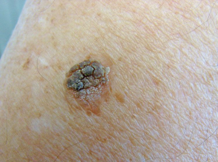
A 59-year-old man became concerned when a long-standing pigmented lesion on the shoulder began to both expand and develop multiple colors.
Key point: The “rough”-appearing surface topography, truncal location, and age of the patient all suggest that this is a routine seborrheic keratosis.
Treatment: A superficial shave excision/biopsy was done to alleviate the patient’s anxiety. The lesion did prove to be a seborrheic keratosis.
Note: Many seborrheic keratoses are multi-colored. Many will also grow larger after a long period of stability. When you are in doubt, or when the patient is very anxious, a biopsy (or conservative removal) is indicated.


