- Clinical Technology
- Adult Immunization
- Hepatology
- Pediatric Immunization
- Screening
- Psychiatry
- Allergy
- Women's Health
- Cardiology
- Pediatrics
- Dermatology
- Endocrinology
- Pain Management
- Gastroenterology
- Infectious Disease
- Obesity Medicine
- Rheumatology
- Nephrology
- Neurology
- Pulmonology
Rhinocerebral Mucormycosis in a 67-Year-Old Woman
Mucormycosis, an angioinvasive yeast infection of the Mucorales order of the class of Zygomycetes, often grows in patients with diabetes mellitus, especially in the presence of diabetic ketoacidosis.

A 67-year-old woman presented to the emergency department (ED) with delirium resulting from diabetic ketoacidosis. She also complained of headache, facial pain, nasal congestion, left eye pain, and blurry vision of 2 weeks’ duration. She noted recent bloody nasal discharge and confusion, which was her reason to come to the ED.
The patient’s past medical history was significant for type 2 diabetes mellitus (DM) with neuropathy, arthritis, multiple urinary tract infections, cataracts, and obesity. She denied smoking and alcohol and drug use. She lives alone with a cat.
Vital signs were temperature, 36.7°C (98°F); pulse, 114 beats/min; respiratory rate, 25 breaths/min; blood pressure, 116/20 mm Hg; oxygen saturation, 98% on room air. She was not oriented to place or time.
Examination revealed signs of inflammation and some exudates in the patient’s left ear canal, dried blood around her nares, and dry mucous membranes.
Lung and heart examination findings were normal.
Laboratory findings were significant: fasting glucose level of 800 mg/dL (normal range, 70 to 100 mg/dL), with an anion gap of 28 mmol/L (normal range, 2 to 16 mmol/L) and hemoglobin A1C (HbA1c) level of 15.9% (normal range, 5% to 6%). The leukocyte count was 32.3/mL (normal range, 4.5 to 10/mL), with 7% bands (normal range, 3% to 5%).
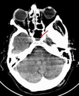
A CT scan of the patient’s sinus (Figure 1, see above) showed ethmoidal sinus congestion with air-fluid levels in the sphenoidal sinuses. Maxillary sinuses and turbinates appeared thickened, and there was left supraorbital soft tissue swelling.
The patient was admitted to the ICU and placed on a diabetic ketoacidosis protocol. By the third day after admission, the diabetic ketoacidosis had resolved; however, the patient continued to complain of sinus tenderness and headache. Otolaryngology debrided her sinuses, and this decreased her pain.

On day 8, the patient was discharged to a nursing facility for general debility and received an outpatient follow-up referral to ENT. During the visit, further sinus debridement showed necrotic areas of the sinuses.
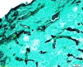
The patient was readmitted and had bilateral maxillary antrostomy, total ethmoidectomy, and sphenoidotomy. Histopathology using Gomori methenamine-silver nitrate stain revealed broad-based, irregular, ribbon-like, nonseptate hyphae with branching that may occur at right angles, as seen in Figure 2, (image to the right).
What’s Your Diagnosis?
Answer and Discussion on next page...
Answer: Rhinocerebral Mucormycosis
Histopathologic examination of multiple tissue samples was positive for Zygomycetes. A diagnosis of rhinocerebral mucormycosis was made.

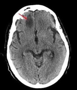
The infectious disease consultant started therapy with IV amphotericin B. A CT scan of the head revealed a hypodense area on the right frontal lobe of the brain (Figure 3, see above).
The patient declined surgical debridement of the brain lesion. A CT scan of the brain obtained weeks later showed near resolution of the brain lesion. The patient was discharged to long-term care to complete 6 weeks of IV amphotericin B therapy. A CT scan obtained 5 months later showed resolution of the frontal brain lesion.
Discussion
Mucormycosis, an angioinvasive yeast infection caused by fungi of the Mucorales order of the class of Zygomycetes, often occurs in patients with DM, especially in the presence of diabetic ketoacidosis. Mucormycosis may be the first manifestation of undiagnosed DM.1 Rhinocerebral mucormycosis is the most common form of mucormycosis in patients with DM.2
Epidemiology. An estimated 500 cases of mucormycosis occur in the United States each year.3 The incidence of mucormycosis appears to have increased over the past 2 decades, with rising numbers of immunocompromised persons and patients with uncontrolled DM.
Pathogenesis. Mucormycosis is associated with uncontrolled DM in 36% to 88% of cases.4 Predisposing factors include elevated blood sugar levels, elevated serum iron levels, an immunocompromised state, hemochromatosis, deferoxamine therapy, hematological malignancies, neonatal prematurity, malnutrition, prolonged corticosteroid therapy, and prophylactic antifungal agents.
Mucormycosis is thermotolerant, and the fungi can be isolated in large numbers from soil or decomposing organic matter, such as bread, leaves, and fruits. Most infection is exogenous and occurs after inhalation of spores.5
Diagnosis. Early diagnosis of mucormycosis is challenging but vital because delayed treatment clearly worsens outcomes. Persons in whom rhinocerebral disease suspected after history and physical examination should undergo emergent CT scanning of the paranasal sinuses and an endoscopic examination of the nasal passages with biopsies of any suggestive lesions. As seen in the CT scan of this patient’s head (Figure 1), she has general swelling of the turbinates with maxillary congestion and air-fluid level on the ethmoidal sinus.
Histopathologic and microbiologic examination of biopsy specimens helps confirm the diagnosis. Gomori methenamine-silver nitrate stain (Figure 2) shows broad-based, irregular, ribbon-like, nonseptate hyphae with branching that may occur at right angles.
The differential diagnosis included rhinosinusitis, acute bacterial sinusitis, allergic rhinitis, migraine headache, nasal polyps or tumors, and meningitis.
Management
Treatment of patients with rhinocerebral mucormycosis is based on rapid reversal of the underlying cause, such as metabolic acidosis; antifungal therapy with amphotericin B; and surgical intervention. Timely identification and management of mucormycosis is important for preventing its spread, which may be hematogenous or contiguous. Tacrolimus and statins have been noted to decrease the incidence of mucormycosis because of their activity against Zygomycetes.6
Prognosis
Mucormycosis carries an overall mortality rate of 50% to 85% of cases. The mortality rate associated with rhinocerebral disease is 50% to 70% of cases. The mortality rate of disseminated disease approaches 100%.7
Conclusion
Physicians should be aware of specific fungal infections complicating simple conditions such as sinusitis in patients with DM, especially while they have diabetic ketoacidosis. New-onset diabetic ketoacidosis in a patient with type 2 DM with sinus symptoms should be a red flag.
In addition, any patient with DM who has a headache and visual changes is a candidate for prompt evaluation with imaging studies and nasal endoscopy to rule out mucormycosis.
Of note: A black necrotic nasal eschar is the hallmark of mucormycosis, but its absence does not exclude the condition. For patients with rhinocerebral mucormycosis, early diagnosis and treatment are imperative to avoid spread to the brain and other tissues and to prevent poor outcomes.
References:
1. Kontoyiannis DP. Decrease in the number of reported cases of mucormycosis among patients with diabetes mellitus: a hypothesis. Clin Infect Dis. 2007;44:1089-1090.
2. Roden MM, Zaoutis TE, Buchanan WL, et al. Epidemiology and outcome of zygomycosis: a review of 929 reported cases. Clin Infect Dis. 2005;41:634-653.
3. Rees JR, Pinner RW, Hajjeh RA, et al. The epidemiological features of invasive mycotic infections in the San Francisco Bay area, 1992-1993: results of population-based laboratory active surveillance. Clin Infect Dis. 1998;27:1138-1147.
4. Greenberg RN, Scott LJ, Vaughn HH, Ribes JA. Zygomycosis (mucormycosis): emerging clinical importance and new treatments. Curr Opin Infect Dis. 2004;17:517-525.
5. Richardson MD, Shankland GS. Rhizopus, Rhizomucor, Absidia, and other agents of systemic and subcutaneous zygomycoses. In: Murray PR, Baron EJ, Pfaller MA, et al. Manual of Clinical Microbiology. 7th ed. Washington DC: ASM Press; 1999:1242-1258.
6. Reed C, Bryant R, Ibrahim AS, et al. Combination polyene-capsofungin treatment of rhino-orbital-cerebral mucormycosis. Clin Infect Dis. 2008;47:364-371.
7. Spellberg B, Edwards J Jr, Ibrahim A. Novel perspectives on mucormycosis: pathophysiology, presentation, and management. Clin Microbiol Rev. 2005;18:556-569.
